|
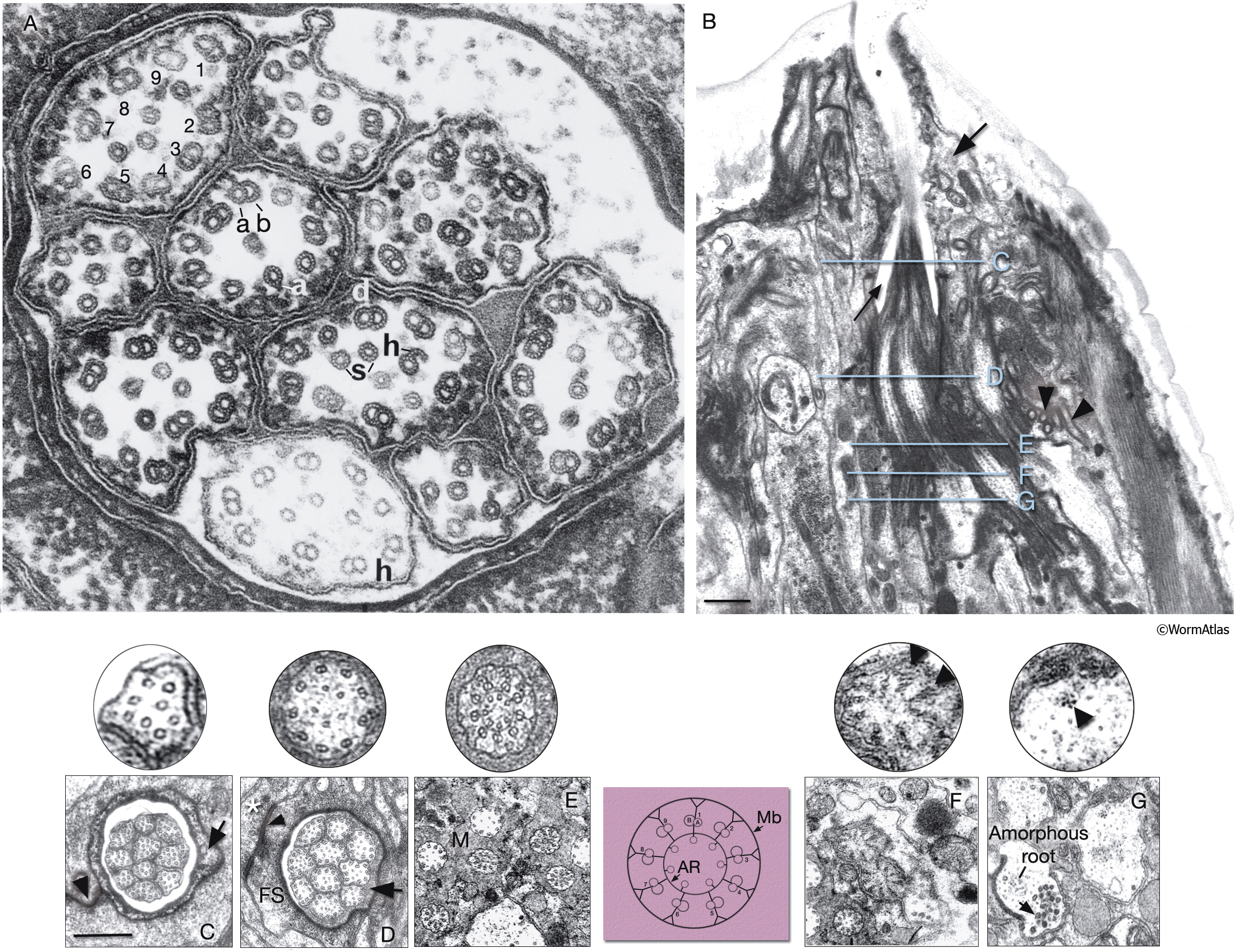
NeuroFIG 23: Ultrastructure of amphid cilia.
A. Ciliated endings of eight amphidial neurons. In this example, many cilia are sectioned through their “middle segment,” transverse section. Each cilium contains 9 MT doublets (d), which are composed of A sub-fibers (a) with 13 protofilaments and B subfibers (b) with 11 protofilaments. Each cilium also possesses a variable number (1–6) of inner singlet 11p MTs (s). Hook (h) structures are seen as the B subfibers end near the end of the middle segment. Distal to the ends of B subfibers, nine 13p MTs are seen around the periphery of the process with a smaller number of 11p singlets inside. Magnification, 300,000x, TEM image. (Reprinted, with permission, from Chalfie and Thomson 1982. ©The Rockefeller University Press.)
B. TEM of the left amphid channel, longitudinal section. (Same section as in NeuroFIG 35B, magnified.) Amphid cilia exposed to the outside are clustered within the amphid channel (thin arrow). The fingers of the AFD neuron embedded within the sheath cell are seen to the right of the channel (arrowheads). The arrow points to the socket cell, which surrounds the distal portion of the channel. The levels of sections in panels C–G are shown with straight blue lines in B. Anterior is at the top of the panel. Bar, 1 μm.
C–G. Transverse TEM sections through the amphid channel at various levels. Bar, 0.5 μm. (Top panels) A single cilium magnified further.. (Based on Perkins et al., 1986.) C. Section through the distal segments, showing ten channel cilia about 3 μm from the tip of the lips. At this level, each cilium contains only the 13p A subfibers, which continue into the distal segment, and several 11p singlet MTs. The cuticle lines the surface of the socket channel (arrow). (Arrowhead) Self-junction of the socket cell (Perkins et al., 1986).
D. Section through the middle segments, about 5.5 μm from the tip of the lips. Nine doublet MTs (with both A and B subfibers) are attached to the membrane and two to seven smaller singlet MTs occupy the center of each cilium (five are seen in the top panel). No apical ring is seen yet. An electron-dense material lines the sheath channel surface and a scant matrix is seen inside this surface surrounding the cilia (arrow). Sheath-cell cytoplasm is filled with a filamentous scaffold (FS) around the channel. A sheath cell makes adherens junctions (arrowhead) to the socket cell (asterisk), which has moved peripherally by this level.
E. Section through the transition zone, which is about 0.27 μm in diameter and 1 μm long in each cilium. Matrix (M) separates the cilia in the channel. Within each cilium, the nine doublets are drawn together by the apical ring and are attached to the cilium membrane by Y-shaped links. The seven singlets are aligned along the inner face of the apical ring and are also attached to it. These inner singlets also originate at the base of the transition zone. (Colored panel) Schematic rendition of the structure of the transition zone. (Mb) Cilium membrane; (AR) apical ring.
F. Section through the transitional fibers (arrowheads, top panel), about 8 μm from the tip of the lips. Transitional fibers, which may contain a residue of the nematode centriole, join the ends of the doublets radially to the cell membrane. Instead of a distinct basal body, an amorphous root is seen in the center of the dendrite.
G. Section posterior to the transitional fiber zone showing two dendrites. The left dendrite has an amorphous root and is filled with coated pits and vesicles (arrow) just proximal to the base of the cilium. The dendrite on the right is from a more proximal level behind the neuron-sheath junction. This dendrite contains a bundle of seven neurofilaments (arrowhead, top panel). The amorphous root, about 1 μm long, is seen in all channel cilia and the wing cilia. As it tapers out, a fascicle of 3–12 neurofilaments, which are a few micrometers long, becomes visible. There is no amorphous root in AFD dendrites.
Click on picture for full resolution image.
|