|
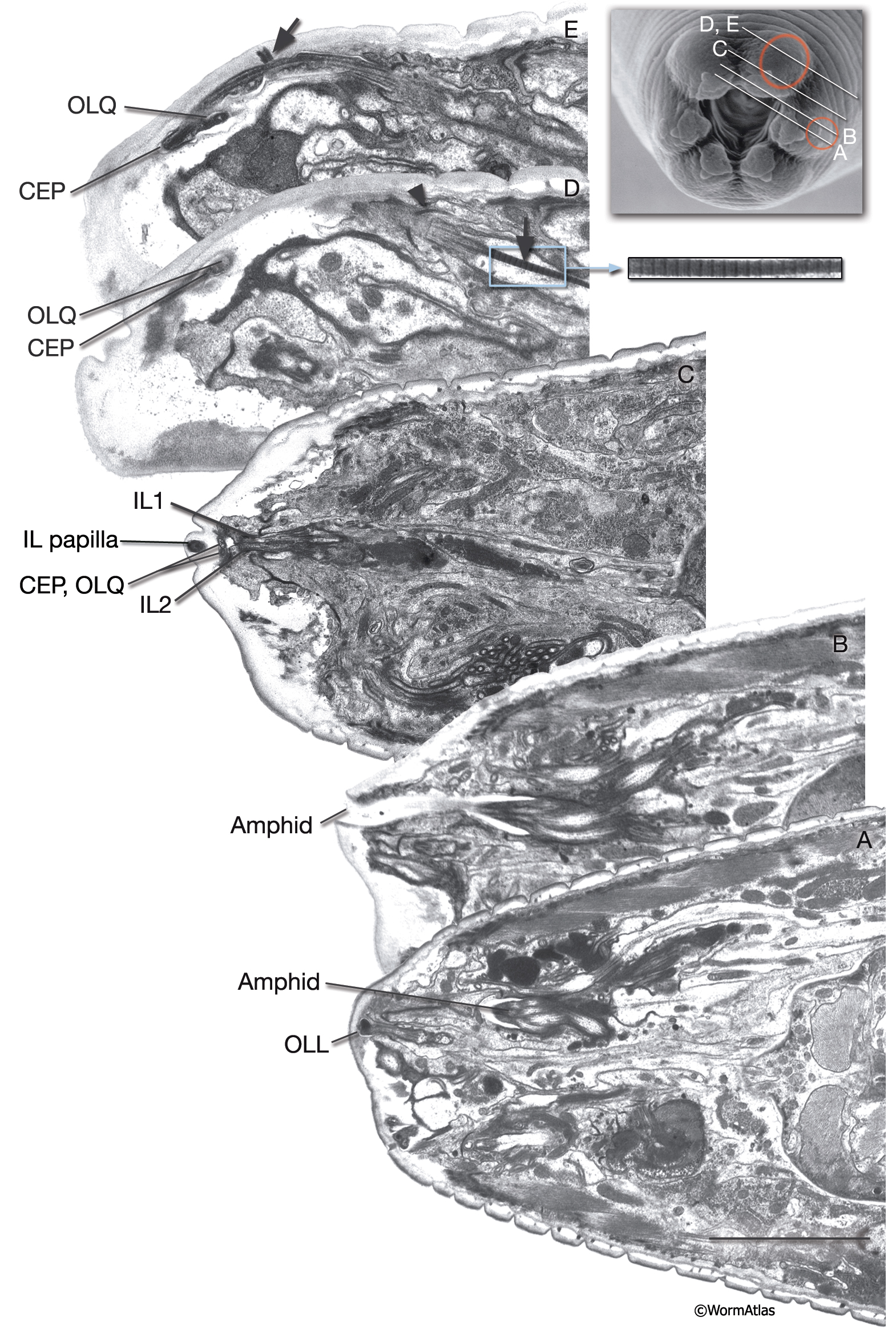
NeuroFIG 35: Ultrastructure of two lips.
The amphid and OLL sensilla have twofold symmetry and reside in the lateral lips. CEP and OLQ sensilla have fourfold symmetry and reside in the dorsal and ventral lips. IL sensilla have sixfold symmetry and are located in all six lips.
A–E. TEMs, longitudinal oblique sections. (Top right inset) Scanning electron microscopy (SEM) image indicating the approximate section levels in A–E. Bar in A, 5 μm.
A. Section through the left lateral lip with the OLLL cilium ending in the cuticle. The many cilia within the left amphid channel can be seen tightly clustered. (Image source: B156 [Hall] 7793.)
B. Section through the left lateral lip at the level of the amphid opening. The amphid cilia become narrower within the amphid channel at their distal ends. (Image source: B156 [Hall] 7805.)
C. Section through the tip of the left dorsal lip. Seen is the electron-dense disc at the tip of DL inner labial papilla. The subcuticular termini of CEPDL and OLQDL are seen below the papilla. (Image source: B156 [Hall] 7897.)
D. Section through a more dorsal level than that in C. Seen are closely neighboring OLQ and CEP cilia in the subcuticle. More proximally, the striated rootlet (arrow) of the OLQDL dendrite is visible (the small inset on the right is magnified from the same area). (Arrowhead) Adherens junction between OLQso and hypodermis. (Image source: B156 [Hall] 7896.)
E. Section through the middle and distal regions of CEPDL and OLQDL sensilla. As it enters the cuticle, CEPDL widens and acquires supernumerary MTs and TAM. A nubbin is seen at the base of its distal region (arrow). (Image source: B156 [Hall] 7905.)
Click on picture for full resolution image.
|