Cell Identification:
Complete Cell List of C. elegans
Nomarski Images for Learning the Anatomy by John Yochem
Part I - HEAD
Pharynx Atlas
Head Neurons:
Head Neuron Nuclei
Hermaphrodite Adult
Hermaphrodite Larva
Male
Part II - BODY
Body Neurons:
Hermaphrodite Ventral Cord Motor Neurons I
Hermaphrodite Ventral Cord Motor Neurons II
Male Ventral Cord Neurons
Hermaphrodite and Male Mechanosensory Neurons
Part III - TAIL
Tail Neurons:
Hermaphrodite Tail Neurons
Male Tail Neurons
Post Cloacal Sensilla and Spicule Nuclei - Rene Garcia
L4 and Adult Male Tail
Tail Non-neuronal Cells:
L4 Male Proctodeum and Muscles
Helpful Notes for Cell Identification by Nomarski Microscopy:
 Click pictures for new window with high resolution image Click pictures for new window with high resolution image
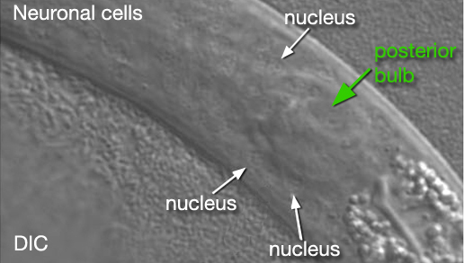
Neuronal nuclei are rather small,
round and have stippled appearance.
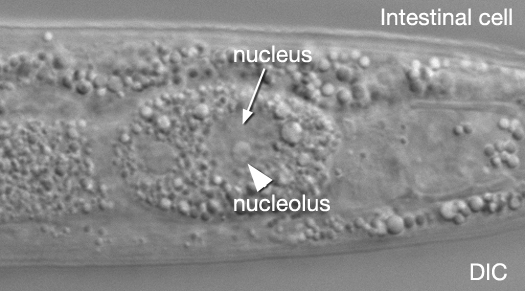
Hypodermal and gut nuclei are
large, have a "fried egg" appearance with large, prominent nucleoli.
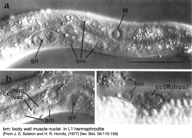
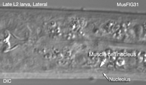
Muscle nuclei are oblong (ovoid), intermediate
in size between neuronal and hypodermal nuclei, and have a small, spherical nucleolus. Their nucleoplasm
is granular in L1 but becomes smooth in L2 and remains so throughout the rest of the development.
Most cells are most easily seen
in very young larvae; L1 stage is optimal for neuronal identifications. Pharyngeal
nuclei are easier to see in the L2 stage.
Some cells are difficult to identify
depending on their cell positions because of natural variability in their location:
the posterior lateral ganglia
of the head (AIN, RIC, AIZ, ADEso, AVD)
the anterior socket and sheath
cells in the head (AMso, ILsh, ILso, OLQso)
postembryonic neurons in the
tail
postembryonic neurons in VC
Ancillary Methods for Cell Identification:
FITC staining , DiI staining, DiO
staining, DAPI staining -->> See Anatomical Methods
References:
Hedgecock E. M. et
al., 1985. Axonal guidance mutants of Caenorhabditis elegans identified
by filling sensory neurons with fluorescein dyes.
Dev. Biol. 111: 158-170.
Bargmann C. I. and
Avery L. 1995. Laser killing of cells in Caenorhabditis elegans. in
Methods in Cell Biology Vol.48,
(ed: Epstein H. F. and Shakes D. C), pp. 225-249. Academic Press, California.
Sulston J. and Hodgkin
J. 1988. Methods in The Nematode Caenorhabditis elegans. (ed: Wood
W. B. and the community of C. elegans researchers),
pp.587. Cold Spring Harbor Laboratory Press, New York.
|
|