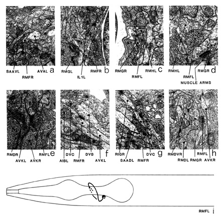
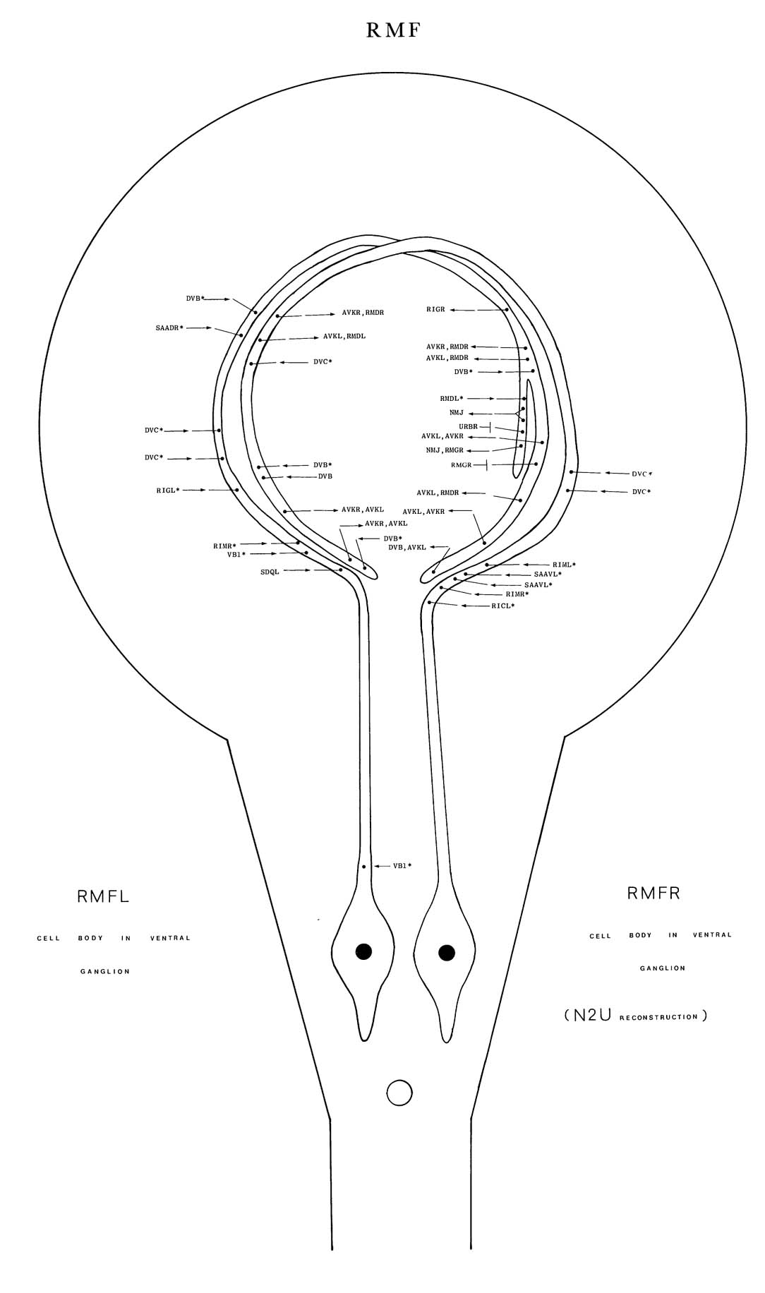
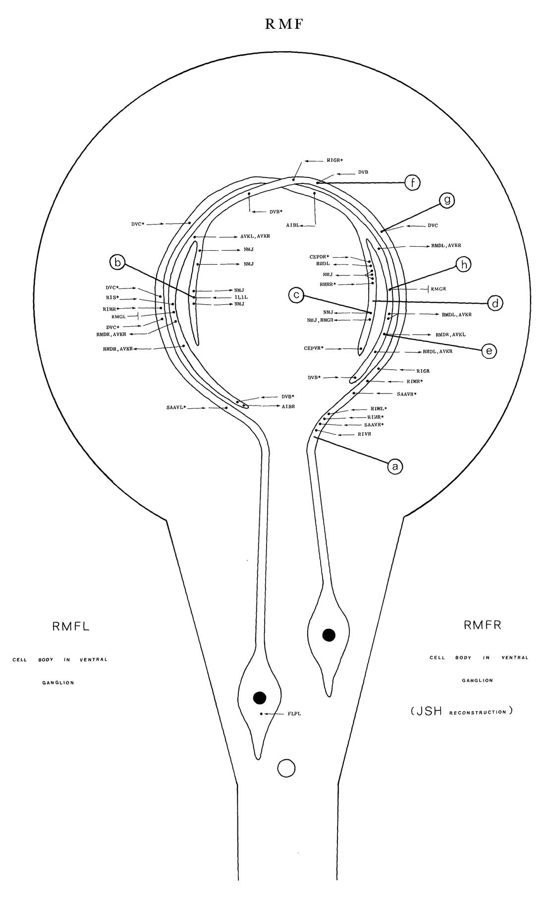
RMF is a set of two motoneurons, which innervate head muscles via NMJs in the nerve ring.
The cell bodies of RMF are situated in the ventral ganglion and send anteriorly directed
processes into the nerve ring. These processes are initially closely associated with those of AVA (a) but then move away from AVA in the nerve ring and run right round it near the outside
surface and anterior face. A branch comes off each process at a distal sub-dorsal position. (Only
one was present in the N2U reconstruction, but two were seen in other reconstructions, so a
diagram of RMF from the JSH animal has been included). This branch moves to the inside
surface of the nerve ring and has NMJs. The main synaptic outputs are: NMJs, which are often
dyadic or triadic with RMG as a corecipient (c); AVK (e); and RMD (e). Some of the synaptic
vesicles of RMF have dark-looking cores (d). The main synaptic input comes from DVB (f)
and DVC (g). There are gap junctions to RMG (h).
Magnifications: (a-c, f-h) x 12750, (d, e) x 25500.


Click pictures for higher resolution images