Click pictures for higher resolution images Click pictures for higher resolution images
PVD is a set of two interneurons with cell bodies situated laterally in the posterior body.
Anteriorly and posteriorly directed processes leave the cell bodies and run alongside the
excretory canal in close association with the processes of ALA and CAN. These three processes
have not been completely reconstructed in the posterior body, although they have been
sampled in several places. Two of the processes end at about the level of the anus; a third enters
the lumbar ganglia and synapses onto PVC (ALA-d). A single synapse on the lateral
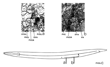
hypodermis has also been seen on one of the processes (CAN-c). PVD neurons send out a third,
ventrally directed process, which enters the ventral cord as a commissure. This process runs
anteriorly in the ventral part of the cord and has several dyadic synapses to AVA and PVC (a). There are also a few synapses to DVA (b).
Magnifications: (a, b) x 25500.
PVD ventral cord synapses
partners |
gap junctions |
synapses from |
synapses to and corecipients |
AVA |
- |
- |
22 PVC, 4AVA, PVD |
PVC |
- |
1m |
2, 22 AVA, 2 PVC, DVA, HDC |
DVA |
- |
- |
2, PVC |
PVD |
- |
1m |
AVA |
HDC |
- |
- |
PVC |

|
|