Click pictures for higher resolution images Click pictures for higher resolution images
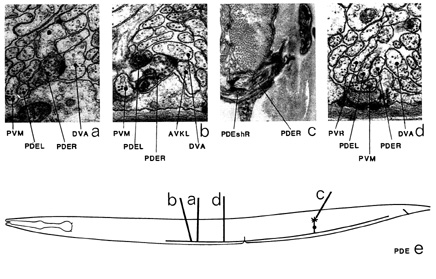
PDE is a pair of neurons with ciliated endings in the posterior deirid sensilla. The cell bodies
of PDE are situated sub-ventrally in the posterior body (e). Dorsally directed processes run
up to the sensilla, which are not in the alae, as are those of ADE, but dorsal to them, next
to the sub-dorsal muscles (c). Ventrally directed processes leave the cell body and enter the
ventral cord via a commissure, where they split and run both anteriorly and posteriorly in the
ventral region of the cord. PDE neurons have been shown to contain dopamine (Sulston et al. 1975). The main synaptic output is to DVA (a, b) and AVK (b); there are also a few synapses
to hypodermal cells. The main synaptic input is from PVM (*g), and there is also some from PLM (*e) and AVK (*c). There are gap junctions to itself (d), PVC and PVM.
Magnifications: (a, b, d) x 25500, (c) x 17000.
PDE ventral cord synapses
partners |
gap junctions |
synapses from |
synapses to and corecipients |
DVA |
- |
1+2m |
37, 20A VK, 2PVR, P DE, PVM |
AVK |
- |
2+2m |
1, 20DVA, HDC |
HDC |
- |
- |
2, AVK, VA9 |
PVC |
- |
1m |
2 |
PVR |
- |
1m |
2DVA |
PVM |
1 |
3+12m |
DVA |
PDE |
3 |
1m |
DVA |
VA9 |
- |
- |
HDC |
VD9 |
- |
- |
- |
PLM |
- |
5m |
- |
AVA |
- |
2m |
- |
AVF |
- |
1m |
- |

|
|