|
MaleRayFIG 2: The ray.
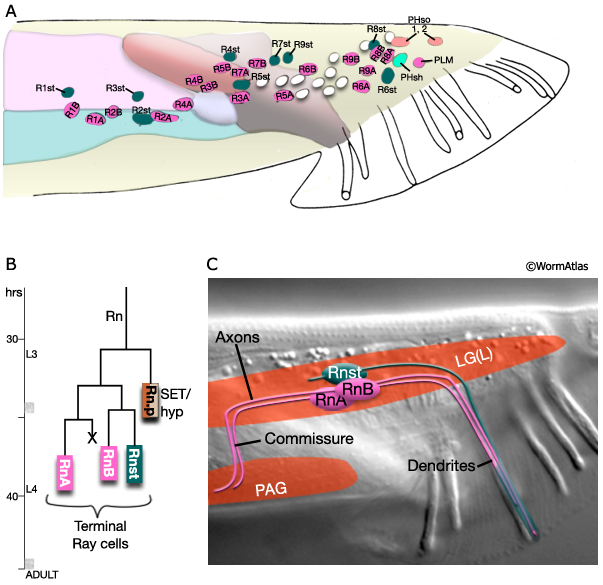
A. Diagram showing nuclei in left lumbar ganglia, lateral view, left side. Ray cell body positions are variable. Unmarked nuclei correspond to juvenile or hermaphrodite cells. (Adapted from Sulston et al., 1980.)
B. Ray sublineage showing cell divisions with time. Hours are post-hatching at 20°C. C. Nomarski DIC with illustration showing ray cell trajectories in the adult ray, lateral view. (Rn) Ray precursor cell; (RnA and RnB) ray neurons; (Rnst) ray structural cell; (PAG) pre-anal ganglion. n=rays 1-9. |