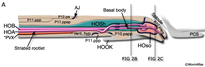
MaleHookFIG 2A: Illustration of the hook.
Cross section of the hook with the locations of the neurons and support cells highlighted, longitudinal view. *PVX is not considered a hook neuron. (Adapted from Sulston et al., 1980.)