|
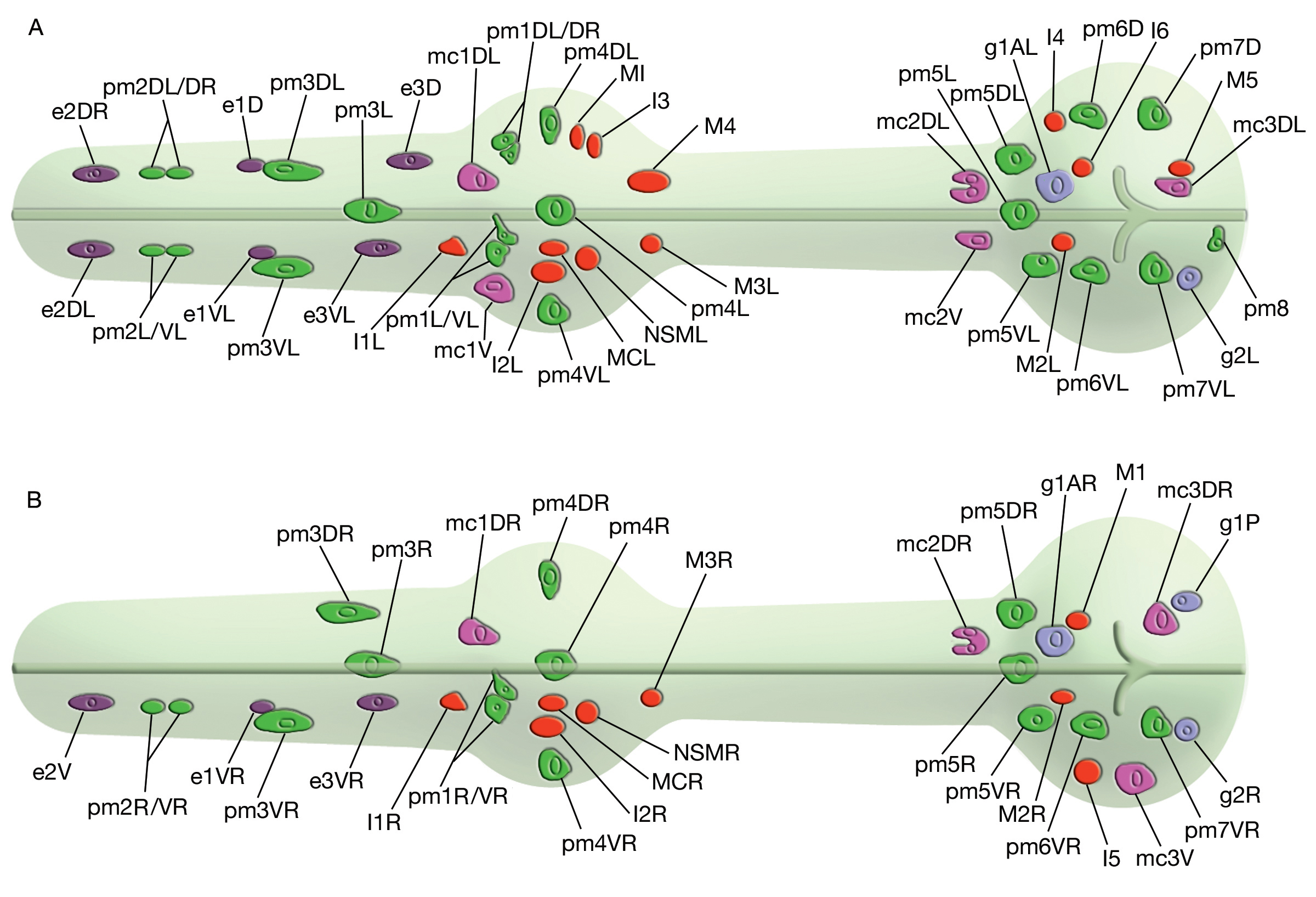
PhaFIG 1: Cell nuclei positions in the pharynx.
(Based on drawing by Ron Ellis.)(Red) Neuron nuclei; (green) pharyngeal muscle nuclei; (lavender) gland nuclei; (fushia) marginal cell nuclei; (purple) epithelial nuclei.
A. Graphic rendition; left lateral view of the ventral, left-side and dorsal nuclei.
B. Graphic rendition; left lateral view of the right-side nuclei.
N.B. More recent studies showed that the identification of g1AL and pm5VL on the left side and g1AR and pm5VR on the right side were switched. This is a corrected image (pers. comm. L Avery).
Click on picture for full resolution image.
|