|
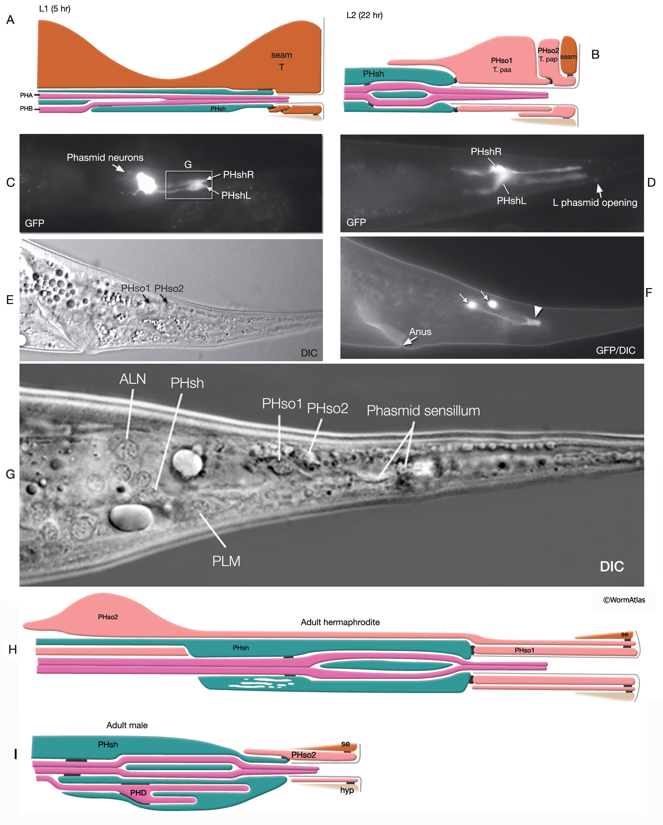
NeuroFIG 41A-I: Structure of the phasmid sensilla.
(Based on Hall, 1977; Sulston et al., 1980.) In the schematic illustrations, dark lines indicate adherens junctions.
A&B. Larval development of the phasmid sensillum. A. Five hours after hatching, the T seam cell functions as the phasmid socket. B. Twenty-two hours after hatching, late-L2 stage. Phasmid structure is still the same between sexes. T-cell daughters T paa (PHso1) and T pap (PHso2) perform the socket cell function. T paa wraps around the channel and is attached to the sheath and T pap by adherens junctions. T pap is attached to the seam and hypodermis by adherens junctions.
C–D. Epifluorescent images from transgenic animals expressing the reporter gene ver-1::GFP in the phasmid sheath cells, left lateral oblique views. DiI-labeled phasmid neurons are also visible in C. Original magnification, 400x. (Strain source: S. Shaham, R. Roubin, and C. Popovici.)
E. DIC image from a transgenic animal expressing the reporter gene T16G1.8::GFP, left lateral view. The two small phasmid socket cell bodies are located slightly dorsally and close to the phasmid sensillum.
F. Same animal, epifluorescent image. Each phasmid socket cells wraps around itself to create a tubular channel (arrowhead) containing the cilia of PHA and PHB neurons. Original magnification, 400x. (Strain source: The Genome BC C. elegans gene expression consortium [McKay et al. 2004].)
G. DIC image of phasmid socket and sheath cells in the tail. Lateral view.
H. Amphid sensillum in the adult hermaphrodite. PHso2 wraps around PHso1, which encircles the channel.
I. Adult male. The socket function is performed by PHso2 while PHso1 transdifferentiates into PHD neuron at L4 (Molina-Garcia et al., submitted, 2019.)
See also NeuroFIG 41J
Click on picture for full resolution image.
|