|
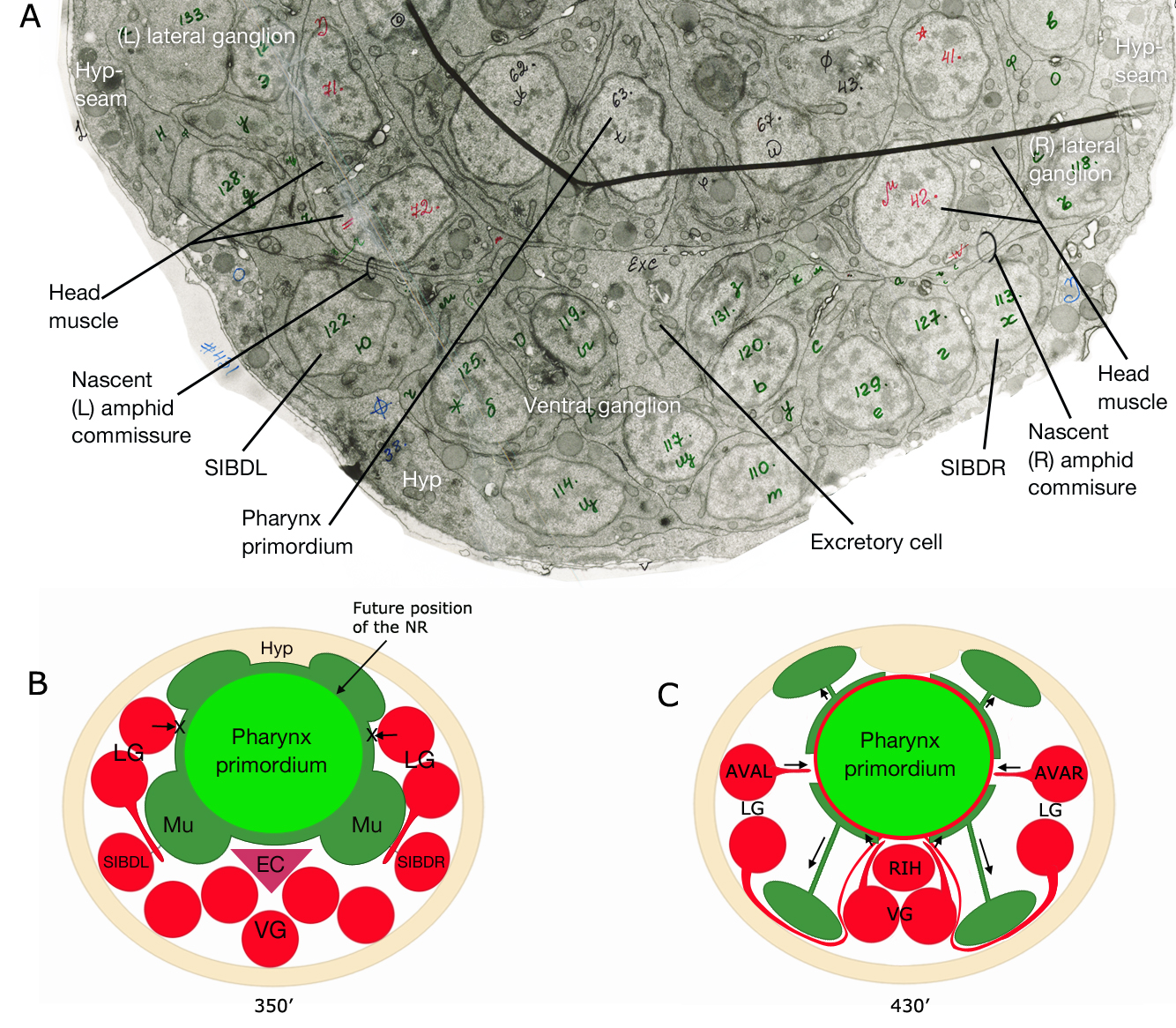
NeuroFIG 15-2: Development of the amphid commissure and the nerve ring (NR).
A. TEM image (cross section) of a 350 min embryo (after first cell division) showing the nascent commissure fibers traveling between the head muscle and the SIBD neuron that has lodged itself between the muscle and the hypodermis (Hyp). B. Schematic representation of the amphid commissure development. 350 min embryo (after first cell division). At this stage muscle cells (mu) that surround the pharynx primordium may be preventing a more direct, i.e. lateral, route to the NR (indicated with crosses) for the processes of the lateral ganglia neurons that are trying to reach the NR. EC, excretory cell; LG, lateral ganglion; VG, ventral ganglion C. Schematic representation of the NR development. 430 min embryo (after first cell division). Muscle cells have migrated to their final positions next to hypodermis, leaving a trailing process behind that remains attached close to the pharynx. Processes from the remaining lateral ganglia neurons can now reach the NR directly from a lateral route rather than having to travel ventrally first. The pioneering neuron to grow a lateral process into the NR is thought to be AVA. Ventrally, RIH is positioned to navigate processes growing into the NR (Norris, C., Hall, D. H., Hedgecock, E. unpublished observations) (Image source: [Hall archive] N611_402.)
Click on picture for full resolution image.
|