|
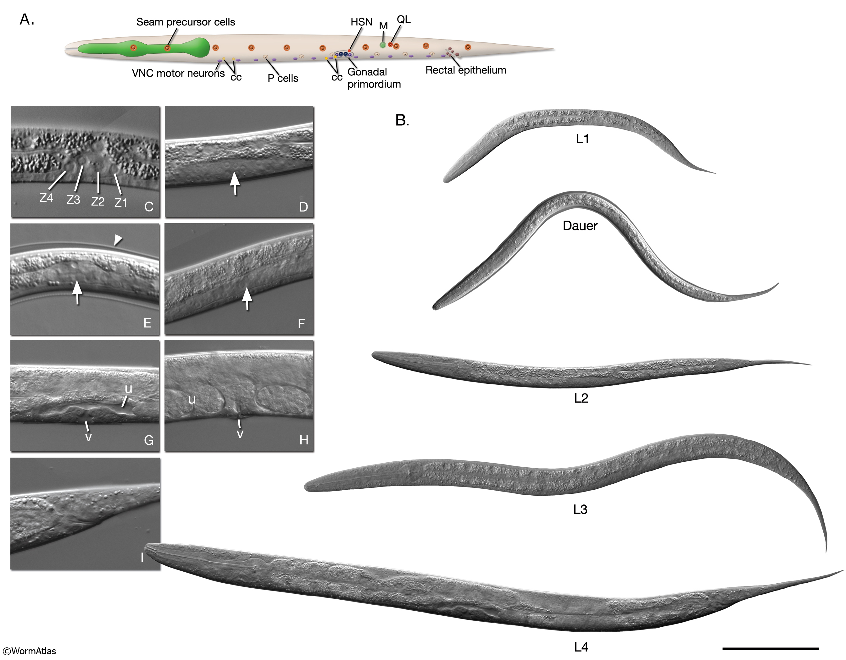
IntroFIG 8: Larval stages of development.
A. L1 larva. The anterior ventral pair of coelomocytes (cc) and the M cell are located on the right. Rectal epithelial cells are in the middle plane, and VNC motor neurons are located at the ventral midline. The remaining cells are as seen from the left lateral side. VNC motor neurons are more numerous than shown. (HSN) Hermaphrodite-specific neuron.
B. DIC images of each stage larva. Bar 0.1 mm.
C- H. Enlarged DIC images of gonads of L1-adult stage animals respectively. Sizes are not to scale. Arrows point to gonads in D-F. (v) Vulva; (u) uterus.
C. Four primordial gonad cells are labeled in L1.
D. Germ cells increase in number in L2.
E. The gonad is similar to an early L2 stage gonad in dauer. Arrowhead points to dauer-specific cuticle.
F. The gonad has extended along the ventral body in L3.
G. Hermaphrodite somatic structures have formed by mid-L4 stage.
H. Vulva is open to the outside and the uterus is full of fertilized eggs in adult.
I. DIC image of L4 tail, which retains ship-like morphology in the hermaphrodite (compare with IntroFIG5E).
Click on picture for high resolution image. |