|
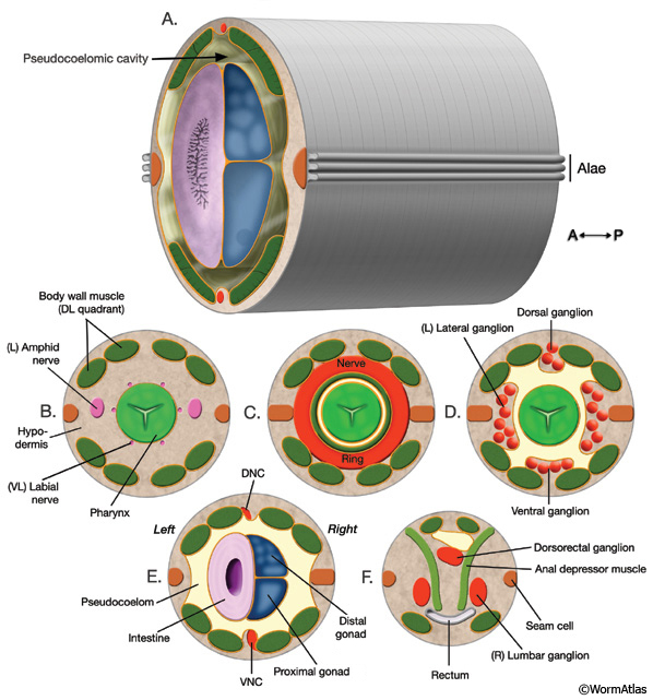
IntroFIG 2: Nematode body plan with cross sections from head to tail.
Approximate level of each cross section is labeled in IntroFIG 1B. Orange lines indicate basal laminae.
A. Posterior body region. Body wall (outer tube) is separated from the inner tube (alimentary system, gonad) by a pseudocoelom.
B. Section through anterior head. The narrow space between the pharynx and the surrounding tissues anterior to the NR can be considered an accessory pseudocoelom because the main pseudocoelom is sealed off by the GLRs at the NR level.
C. Section through the middle of head.
D. Section through posterior head.
E. Section through posterior body.
(DNC) Dorsal nerve cord; (VNC) ventral nerve cord.
F. Section through tail, rectum area.
Click on picture for high resolution image. |