|
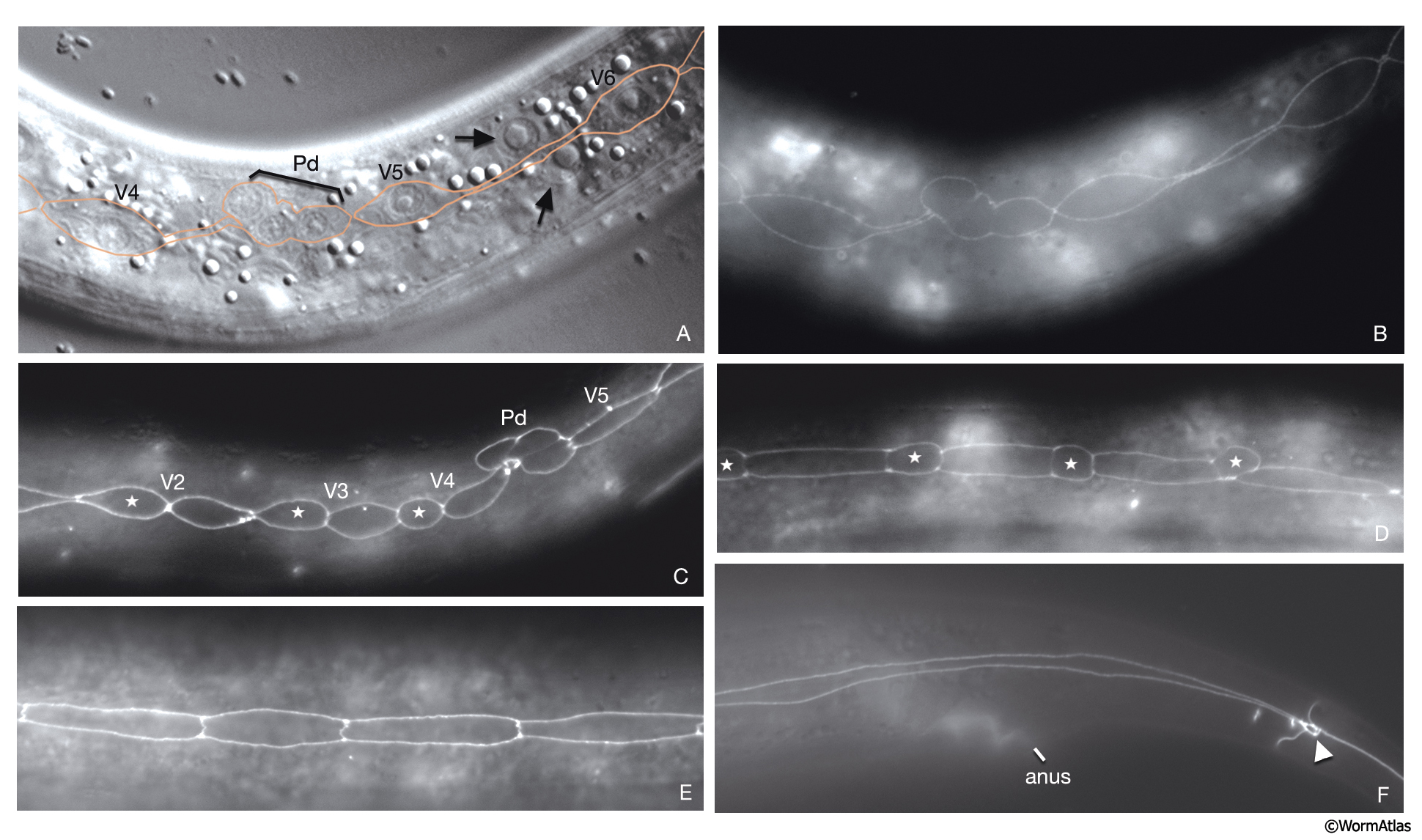
HypFIG 7: Larval divisions of seam cells.
The pattern of division, fusion, and coupled elongation of seam cells repeats itself in each larval stage until mid L4.
A. DIC image of the midbody of an early L2-stage animal, lateral view. The anterior daughters of V5 generate the posterior deirid sensilla (Pd). (Orange line) Seam cells; arrow points to two nuclei of hyp 7.
B-F. Epifluorescent images of animals from a strain expressing the ajm-1::GFP reporter. Original magnification, 600x. (Strain source: H. Yu and P.W. Sternberg.)
B. Epifluorescent image of the same animal as in A. Visible are apical boundaries of dividing seam cells.
C. Epifluorescent image, left lateral view. Shown is the same body region as in A and B, although at a slightly later time in L2 stage. All seam cells have generated daughters (anterior daughters are shown with stars and posterior deirid cells [Pd]) by this time, although none of them have fused with hyp 7 yet.
D. Epifluorescent image, left lateral view. Division of seam cells at L3 stage. Stars indicate anterior daughters that will fuse with hyp 7.
E. Epifluorescent image, left lateral view. Slightly before mid-L4 stage, seam cells have completed their final division, but have not yet fused to each other to make one syncytium. Around mid-L4, each seam cell fuses with its seam neighbors.
F. Epifluorescent image, left lateral view. Young adult animal. The adherens junctions between seam cells have dissolved generating one syncytial seam with 16 nuclei on each side of the animal and spanning the region between phasmid (arrowhead) and hyp 5.
Click on picture for full resolution image.
|