|
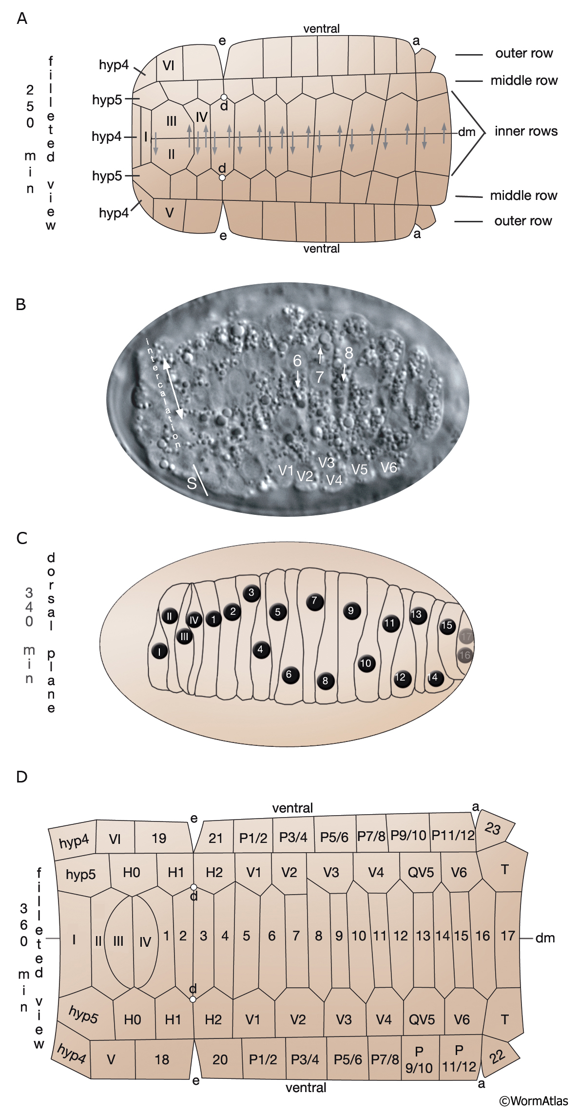
HypFIG 2: Dorsal intercalation of the hypodermis.
A. 250 minutes after first cleavage (filleted view of spheroidal embryo). The hypodermis arises as a patch of cells on the posterior dorsal side of the embryo where the cells organize into six rows. The two inner rows later intercalate (gray arrows) to make the dorsal hypodermis, the middle rows are formed by the seam precursors flanking the dorsal hypodermal cells, and the outer rows contain ventral hyp and P cells. (dm) Dorsal midline; (a) anus;(e) excretory pore; (d) anterior deirid.
B. DIC of an approximately 300-minute embryo (dorsal view). During the initial stages of intercalation, the cells become wedge shaped and their basolateral protrusions insert underneath the adherens junctions of their opposite side neighbors. These basal protrusions then plow through their opposing neighbors while the nuclei of the migrating cells trail behind the basal tips. White arrows point to directions of migration. Nuclei of some hyp 7 cells are labeled for comparison with A and C. (S): Left-side seam progenitors (only V1-V6 are labeled). Two intercalating columns are seen posterior to the double-headed arrow .
C. At 340 minutes (dorsal plane). When the intercalation of dorsal cells is complete, the nuclei are positioned in a dorsolateral location near the border of the seam cells, and new adherens junctions are formed between neighboring cells. The cells have changed from a rounded to an oblong shape. Nuclei 16 and 17 (of hyp 7) are located at the ventral turn of the dorsal hyp. Anterior hyp cells are not shown.
D. At 360 minutes (filleted view). Dorsal hypodermal cells have completed their intercalation that will be followed by ventral enclosure and cell fusions to generate the main body syncytium of hyp 7. Cells I-VI will also fuse to make hyp 6 syncytium. hyp 1-3 cells, one hyp 4 cell (located anterior to IV), and tail hypodermal cells are not shown here (HypFIG 11A). (dm) Dorsal midline. Cells are numbered according to Podbilewicz and White (1994) (cell 1 in these images corresponds to cell 6 in Sulston et al., 1983).
Click on picture for full resolution image.
|