|
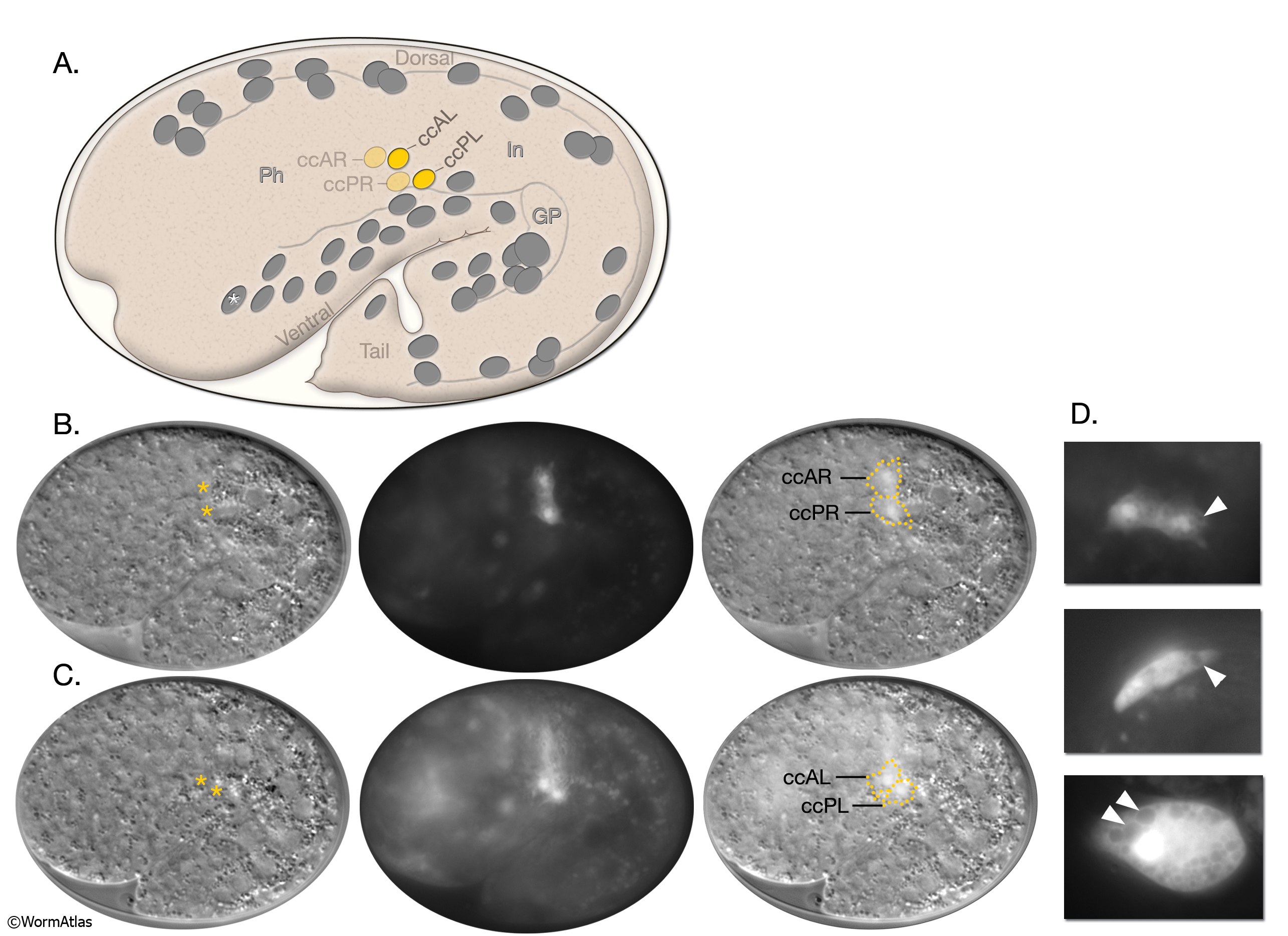
CcFIG 3: Two pairs of coelomocytes are generated in the embryo.
A. The embryonic coelomocytes at approximately 470 minutes after fertilization at 20oC, as seen from the left side. The left- (ccAL and ccPL) and right-side (ccAR and ccPR) coelomocytes are born from symmetric divisions after their mothers migrate posteriorly. The non-motile sister (MSapapap) of the left coelomocyte mother cell, which becomes a body wall muscle, is marked with an asterisk. (Thin gray lines) Borders of the pharynx (Ph), intestine (In), and gonad primordium (GP). (Based on Sulston et al., 1983.)
B. DIC (left), epifluorescent (middle) and DIC/epifluorescent (right) micrographs of a 1.5-fold embryo expressing the reporter gene, unc-122::GFP in the right coelomocyte pair. Asterisks label the nuclei of coelomocytes.
C. The same embryo as in B shown at the left-side level of the animal. The left side coelomocyte pair (ccAL and ccPL) is seen.
D. Epifluorescent images of coelomocytes in 1.5-fold (top), three-fold (middle), and adult (bottom) animals expressing unc-122::GFP. The size of the coelomocytes and the sizes of their endocytic vesicles (arrowheads) increase as the animal grows. Magnification, 600x. (Strain source: D. Williams and E. Jorgensen.)
Click on picture for full resolution image. |