|
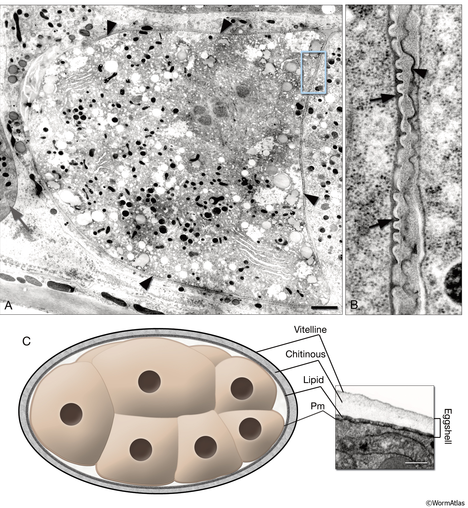
EmbryoIntroFIG 2: Fertilization membrane and eggshell.
A. For a brief period, a recently fertilized embryo becomes covered with a dense, crenelated membrane (arrowheads) that may prevent polyspermy. (Gray arrow) Spermatheca. TEM-high pressure freeze fixation, transverse section. Bar, 1 μm. (Image source: [Hall] CL2099-2A.)
B. Same image as in A, magnified. The fertilization membrane of the embryo on the left (arrows) is still attached to its surface whereas that of an older embryo on the left is being sloughed off (arrowhead).
C. Graphic rendition of the eggshell of an embryo. The eggshell consists of three layers secreted by the egg itself: an outer vitelline layer, a middle chitinous layer, and an inner lipid-rich layer. (Pm) Plasma membrane. The inset on the right is a TEM image of an eggshell. Laser hole fixation. Bar 0.3 μm. (Image source: [Hall] N611-565.)
Click on picture for full resolution image. |