|
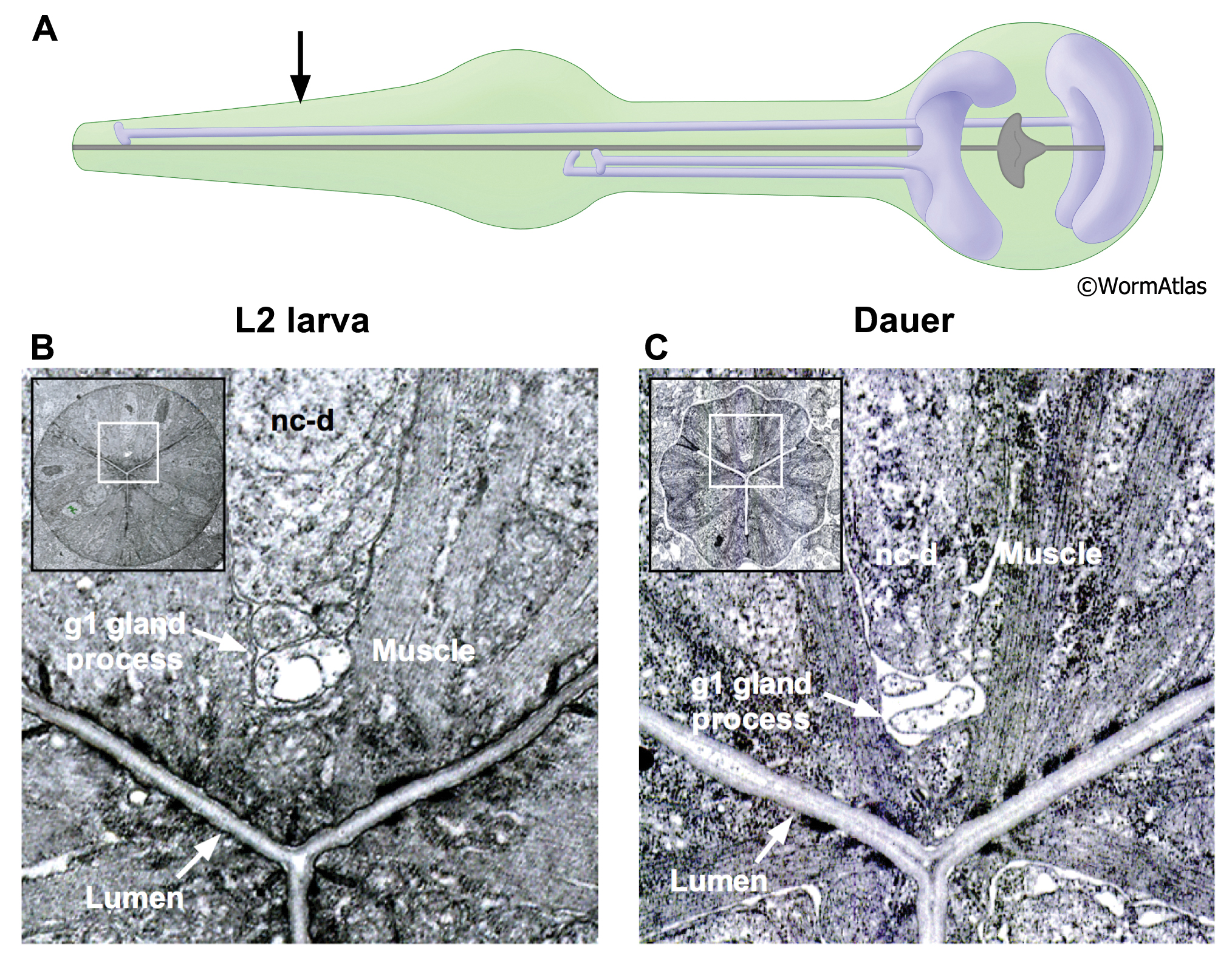
DPhaSUPFIG 5: g1 gland processes.
A. lllustration showing positions of dorsal and ventral g1 gland cells. Arrow indicates approximate position of TEM sections in B&C.
B. Transverse TEM section of L2 larva showing dorsal g1 gland process. (Image source: N2 L2 28-14 [D. Riddle] 750.)
C. Transverse TEM section showing g1 gland process in a dauer larva. Contents of g1 process appears to be more empty and the process diameter is shrunken in the dauer compared to the L2 larva.
Insets, boxed areas indicate regions in enlarged view; nc-d, dorsal pharyngeal nerve cord. (Image source: N2 starved dauer 50-2-1 [D. Riddle] 225M.)
Click on picture for full resolution image.
|