|
DPhaSUPFIG 3: Posterior metacorpus region of dauer pharynx.
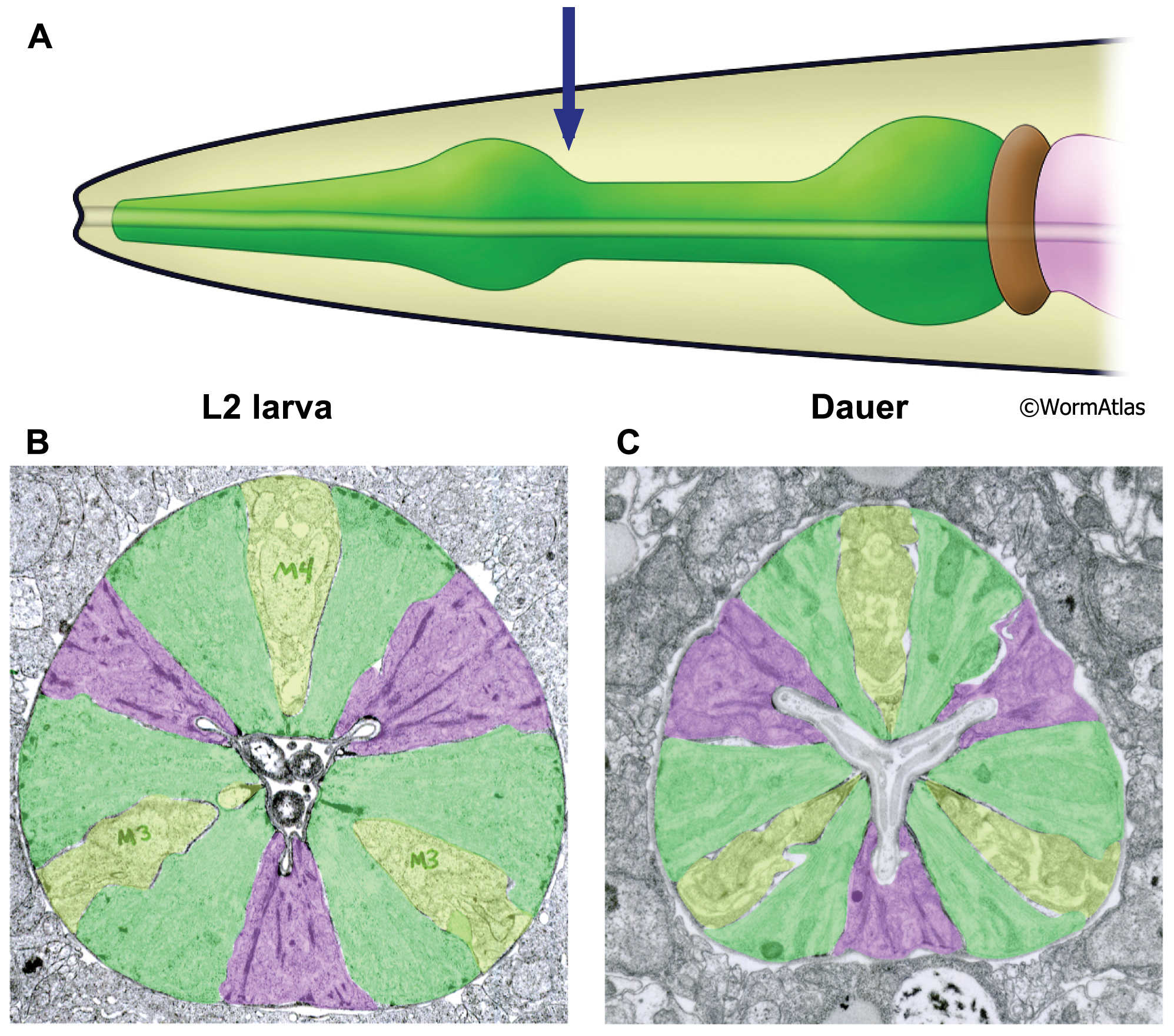
A. Illustration of pharynx with arrow to indicate location of TEM sections.
B. Transverse TEM section of posterior metacorpus in an L2 larva. (Image source: N2 L2 28-14 [D. Riddle] 801.)
C. Transverse TEM section of posterior metacorpus of a dauer pharynx. (Image source: N2 starved dauer 50-2-2 [D. Riddle] 556M.)
B&C. Shading indicates cell types: green, muscle; purple, marginal cells; yellow, nerve cords containing neuronal and gland cells. Markings in B indicate M3 and M4 neuron cell bodies of three pharyngeal neurons in the metacorpus. Images are not presented at the same scale, and were differentially magnified to show structural features.
Click on picture for full resolution image.
|