|
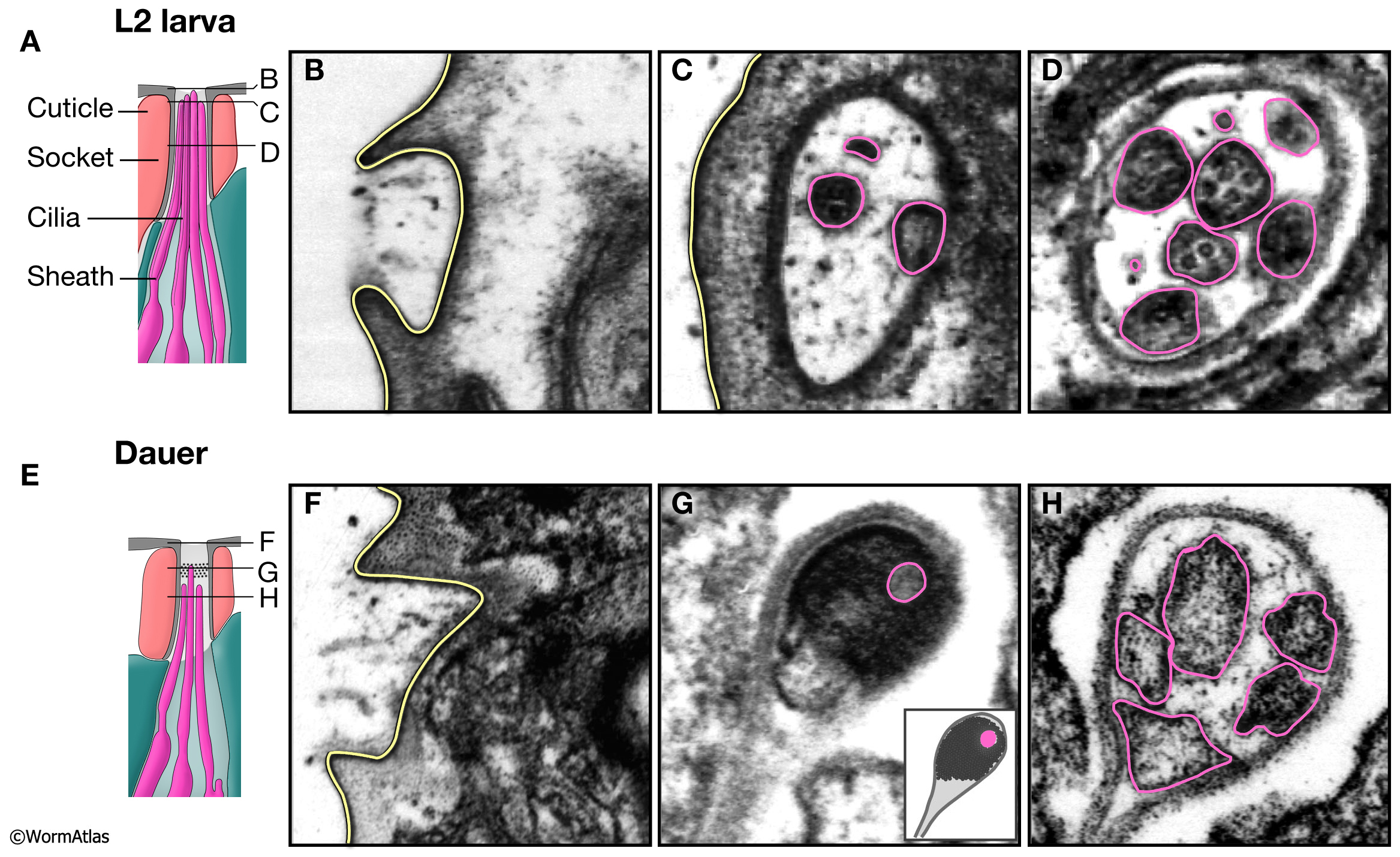
DNeuroFIG 2: Amphid channel openings in L2 and dauer larvae.
A & E illustrate longitudinal views of the amphid channel openings in L2 (A) and dauer (E) larvae.
B-D & F-H show transverse TEM sections whose positions are indicated by horizontal lines in A & E. In L2 and dauer larvae, the amphid channels penetrate the cuticle and are open to the environment (B & F). Yellow lines indicate channel structures contacting the exterior, highlighting an inward dimpling of the lip cuticle where the channel opens to the side of the head (see DNeuroFIG 1).
C,D & G,H show the appearance of cilia tips (pink outlines) within the amphid channels.
G. Electron-dense material fills much of the dauer amphid channel at a level just distal to most cilia tips. Pink shows the position a cilia tip which has penetrated this material. Inset, cartoon illustration of TEM image.
D & H. Cilia in L2 and dauer larvae are similar in appearance in the channel below the level of the electron-dense material.
(Image source: N2 L2 49B [D. Riddle] 19, 22 & 29; N2 starved dauer 50-2-1 [D. Riddle] 33, 42 & 47.)
Click on picture for full resolution image.
|