|
DIntFIG 3: Features in the dauer intestinal cell cytoplasm.
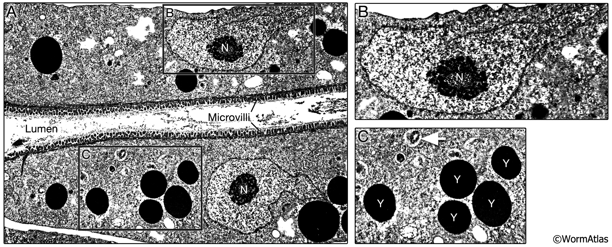
A. Longitudinal TEM photomicrograph of dauer intestinal cell. Dauer intestinal cells contain electron-dense endosomes and yolk granules, probably derived from maternal yolk that was resorbed into the early larval intestine at the end of embryogenesis via the pseudocoelom. The numbers of these yolk granules are substantially fewer in dauers than in L2 or L2d stages. Small autophagosomes appear to be present in dauer intestinal cells, although the contents have not been described.
B. Intestinal nuclei contain an abundance of heterochromatin, with relatively little or no electron-dense euchromatin (condensed chromatin) surrounding a very large nucleolus. As in other stages, the intestinal nuclei are larger than those in other tissues. (Image source: [D. Riddle] N2 dauer 56-6 #109.)
C. Higher magnification view of electron dense yolk granules (Y) and possible small autophagosomes (white arrow) in the dauer intestinal cytoplasm.
Click on picture for full resolution image.
|