|
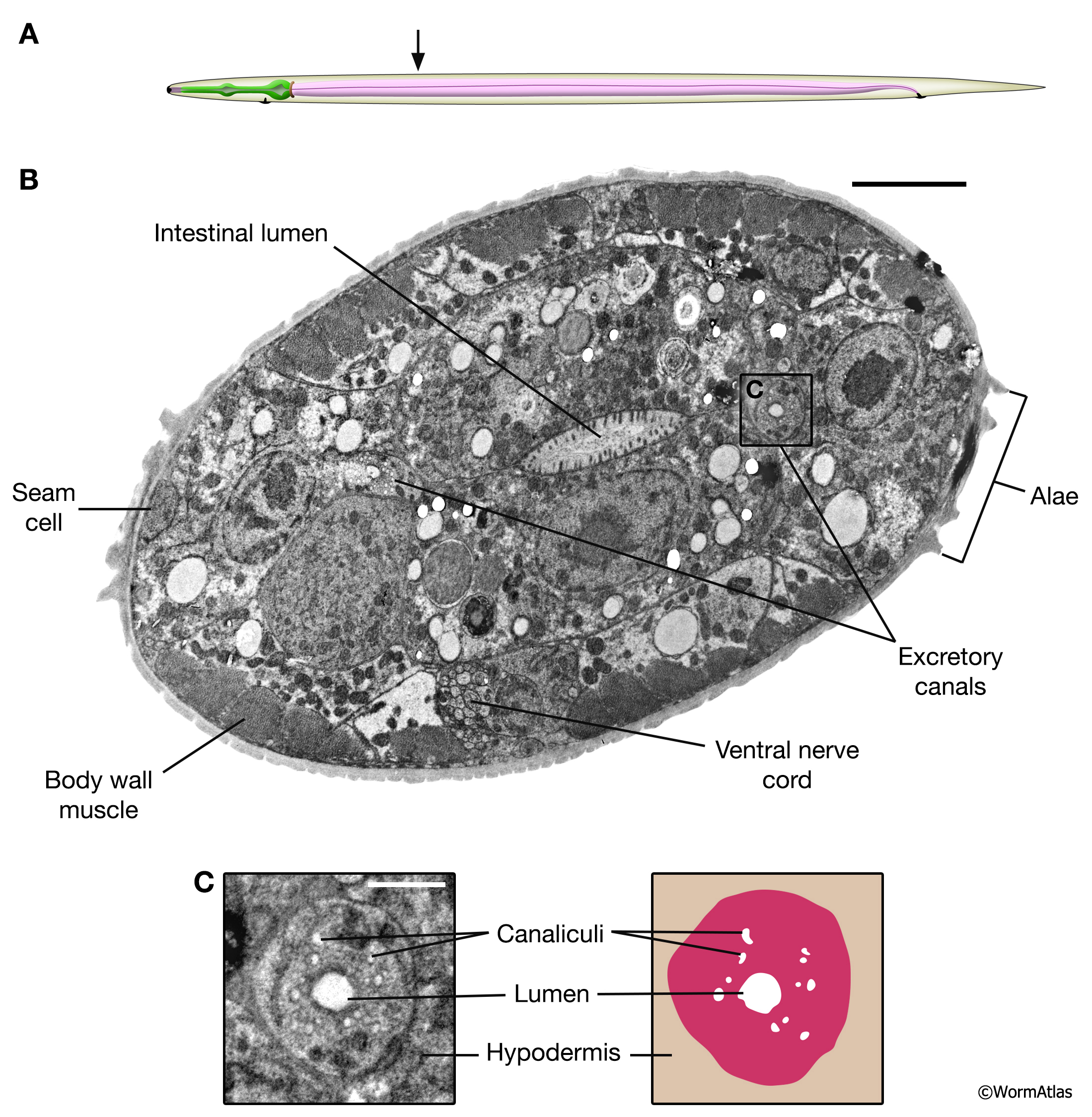
DExcFIG 5: Excretory canal structure in a dauer larva.
Cross-section view from a dauer larvae in the midbody region highlighting bilaterally symmetric excretory canals.
A. Cartoon showing approximate position of section within the dauer body.
B. Transverse, slightly oblique, section showing the midbody region of a dauer larvae. In the midbody, bilateral excretory canals are positioned adjacent to the seam cells, which are easily identified by the dauer cuticular alae. Positions of the intestinal lumen, body wall muscle and ventral nerve cord are also identified. Scale bar, 5 microns.
C. Left panel shows an enlarged view of the excretory canal in cross-section, in which the circular lumen and canaliculi of the canal cell are visible. White arrowheads indicate positions of canaliculi in micrograph. The right panel shows a cartoon representation of the canal cell and internal components. Scale bar, one micron. (Image source: [D. Hall] him-5dar_1127-1_74535_B5_029.)
Click on picture for full resolution image.
|