|
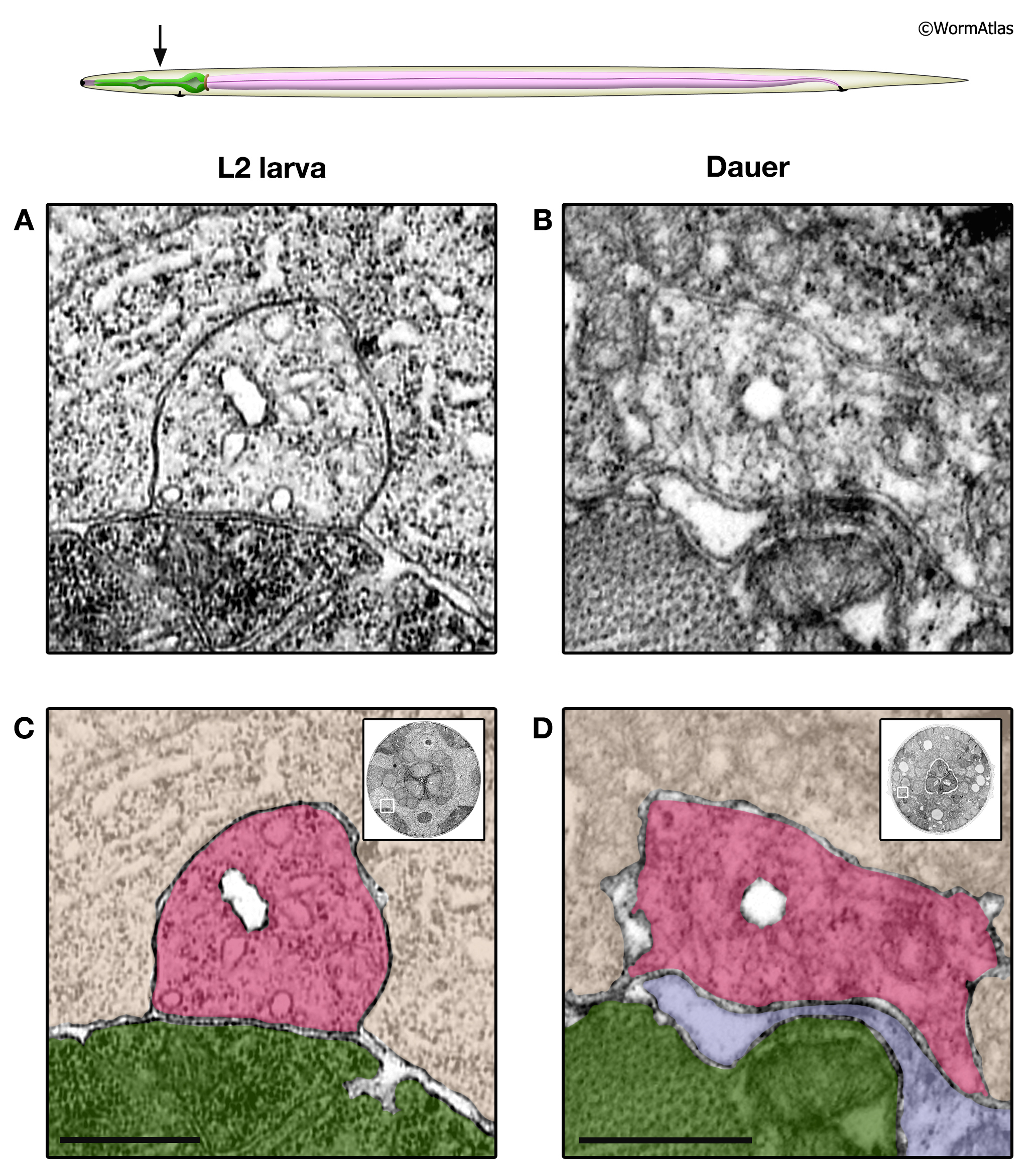
DExcFIG 4: Head regions of excretory canal cells in L2 and dauer larvae.
A. Upper panel, cartoon of approximate location of transverse photomicrographs from the larval head regions in panels A-D.
A&C. Transverse views of excretory canal cells from an L2 larva (A) and dauer larva (B). The excretory canal lumen appears clear, with an apical membrane that is always specialized by an electron dense band of material on its cytoplasmic surface. The several small canaliculi show a simpler single membrane bound, without any special density.
B&D. Images from A&B with shading to indicate different cells. Pink, excretory canal cells with multiple canaliculi; tan, hypodermis; green, muscle; purple, neurons (in B). Inset shows placement of enlarged region within the body section (white box). Bars, 1 micron. (Image sources: L2 [D. Riddle] L2 28-14 #987; dauer [D. Riddle] 50-2-1 #383M.)
Click on picture for full resolution image.
|