|
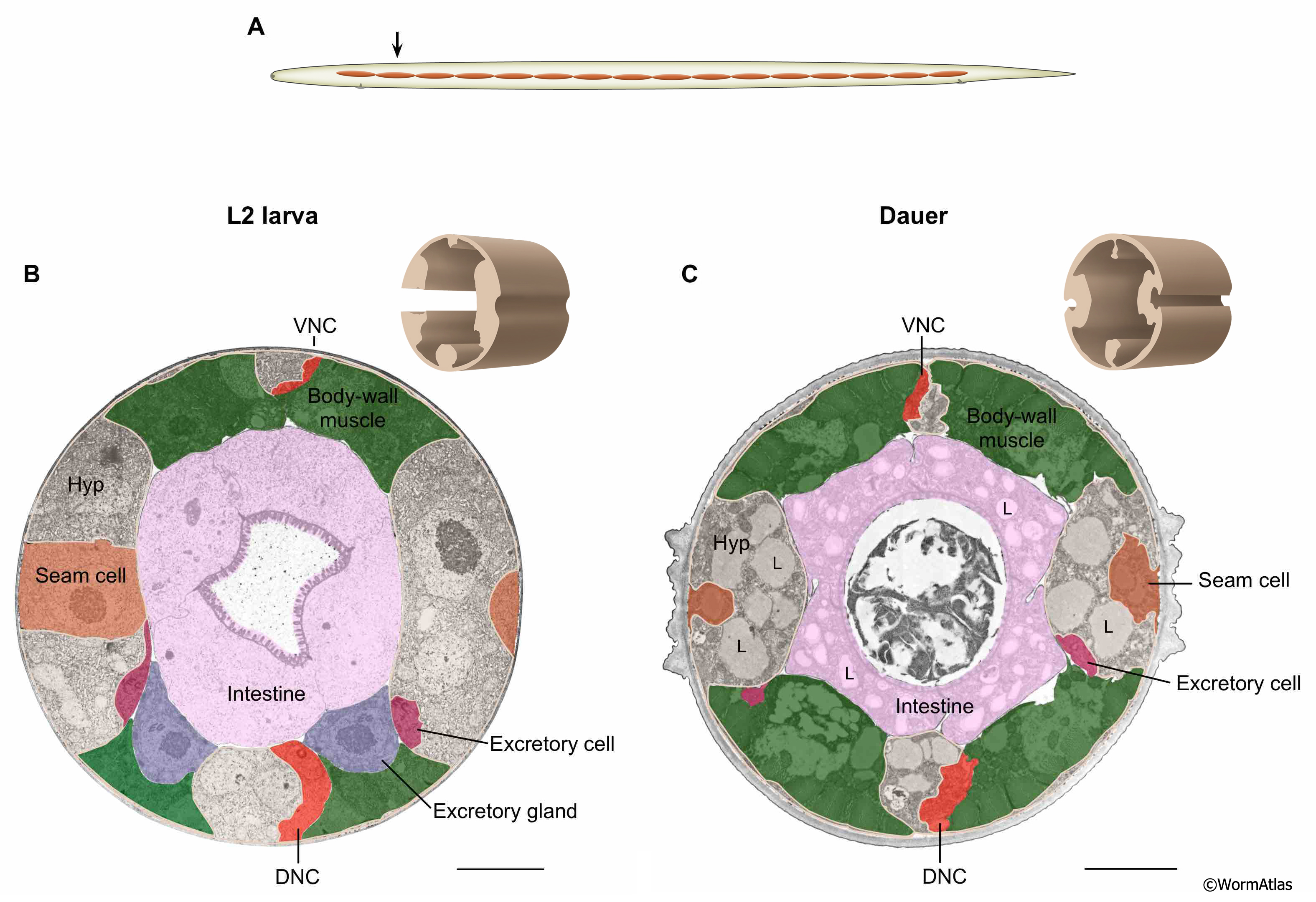
DEpiFIG 1: Hypodermal tissue is shrunken and filled with lipid in dauer larvae.
A. Illustration showing approximate position of sections in lower panels.
B&C. Transverse sections from the anterior intestinal areas of L2 and dauer larva, respectively. 3D rendering of hypodermis from sections in brown shows footprint of tissue. Hypodermis (unshaded) occupies a smaller fraction of the body in the dauer than in the L2 with all four hypodermal quadrants strongly affected. In contrast, the body wall muscle appears roughly similar in size in L2 and dauer larvae (green, see Dauer Muscle section). In dauers, hypodermal shrinkage is due to autophagy (self digestion) (Meléndez et al., 2003). Note the presence of prominent lipid droplets (L) in the dauer hypodermis. VNC, ventral nerve cord; DNC, dorsal nerve cord; Hyp, hypodermis. Scale bar, 5 microns. (Image source: L2 [D. Riddle] N2 28-14 1627; dauer [D. Riddle] 50-2-1 1362.)
|