|
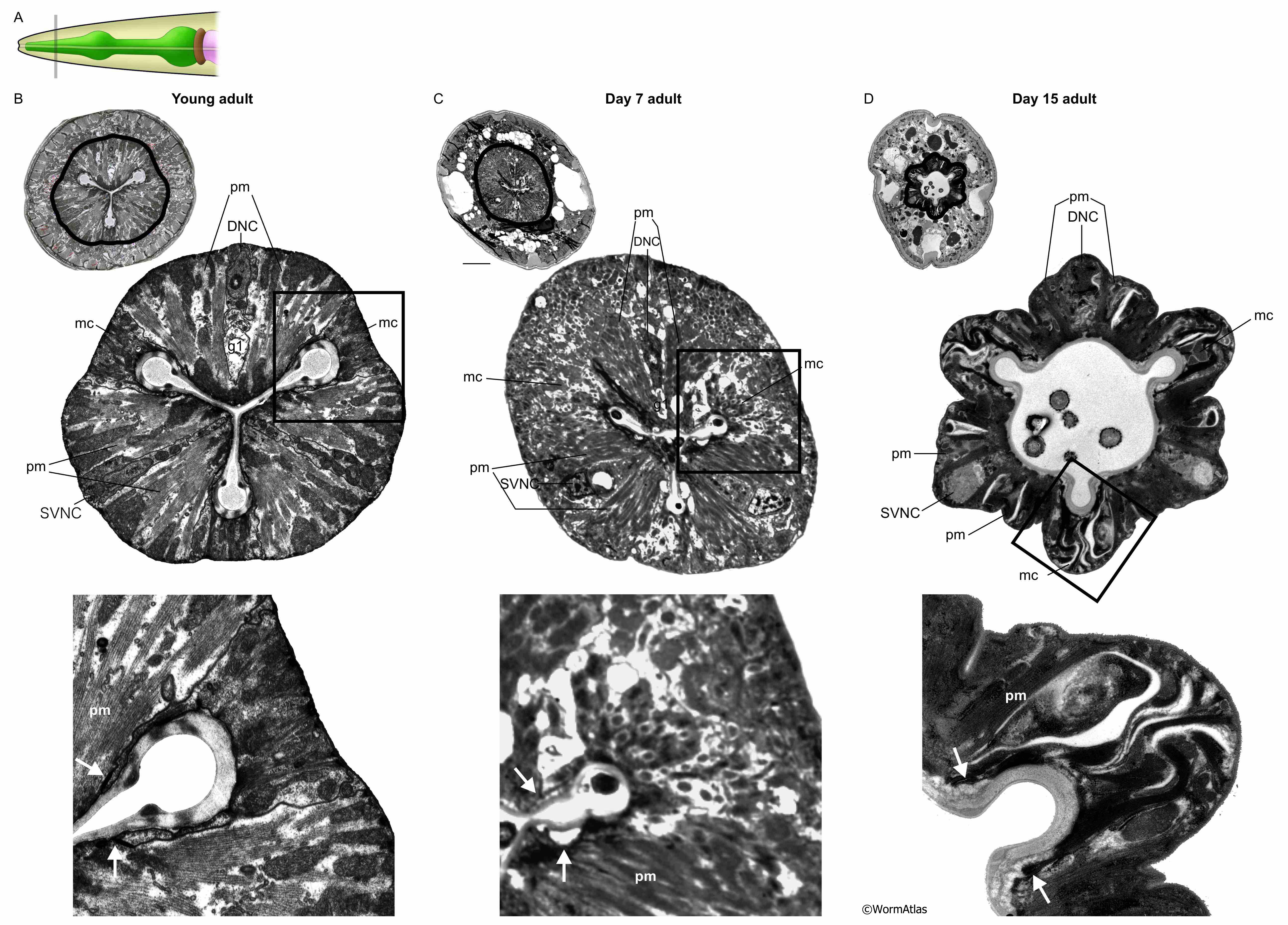
APhaFIG 7: Aging of the procorpus of the pharynx.
A. Diagram of the C. elegans head. Bar shows approximate region of the images in B,C&D.
B. Young adult. C. 7-day-old adult. D. 15-day-old adult.
For B,C&D, upper panels show cross-section of entire head with pharynx outlined with thick black lines. The aging pharynx appears shrunken relative to the head width, occupying a smaller proportion of the head. Panel D is from slightly more anterior, which partially explains the thinner profiles of all anterior pharyngeal tissues here. But the overall shrinkage of the pharynx at 15 days is remarkable, and tissue boundaries are becoming very distorted by un-even shrinkage. Middle panels show the pharynx in greater detail. Lower panels, closeups of the boxed regions in middle panels. In the older animals, the pharynx muscles (pm), marginal cells and nerve cords become more disorganized and the cell boundaries become less well-defined and irregular. White spaces appear between muscle and marginal cells, which may indicate weakened cell attachments. Voids in the muscle and marginal cell bodies may result from cellular deterioration. In the lower panels, marginal cells have detached from the lumen cuticle (white arrows compare similar sites: adherens junctions between pm and marginal cells, and/or, filament attachment zones in the aging animal). The three nerve cords of the pharynx maintain their overall positions but become increasingly disorganized with age. Cells are labeled as indicated: mc, marginal cell; pm, pharynx muscle; DNC, dorsal nerve cord; g1, g1 gland process; SVNC, subventral nerve cord. C, upper panel, scale bar is 5 microns. (Image sources: B, N513A [D. Hall] 1060; C, N826 [D. Hall] G5729; D, N813 [D. Hall] G485 and G488.)
Click on picture for full resolution image.
|