|
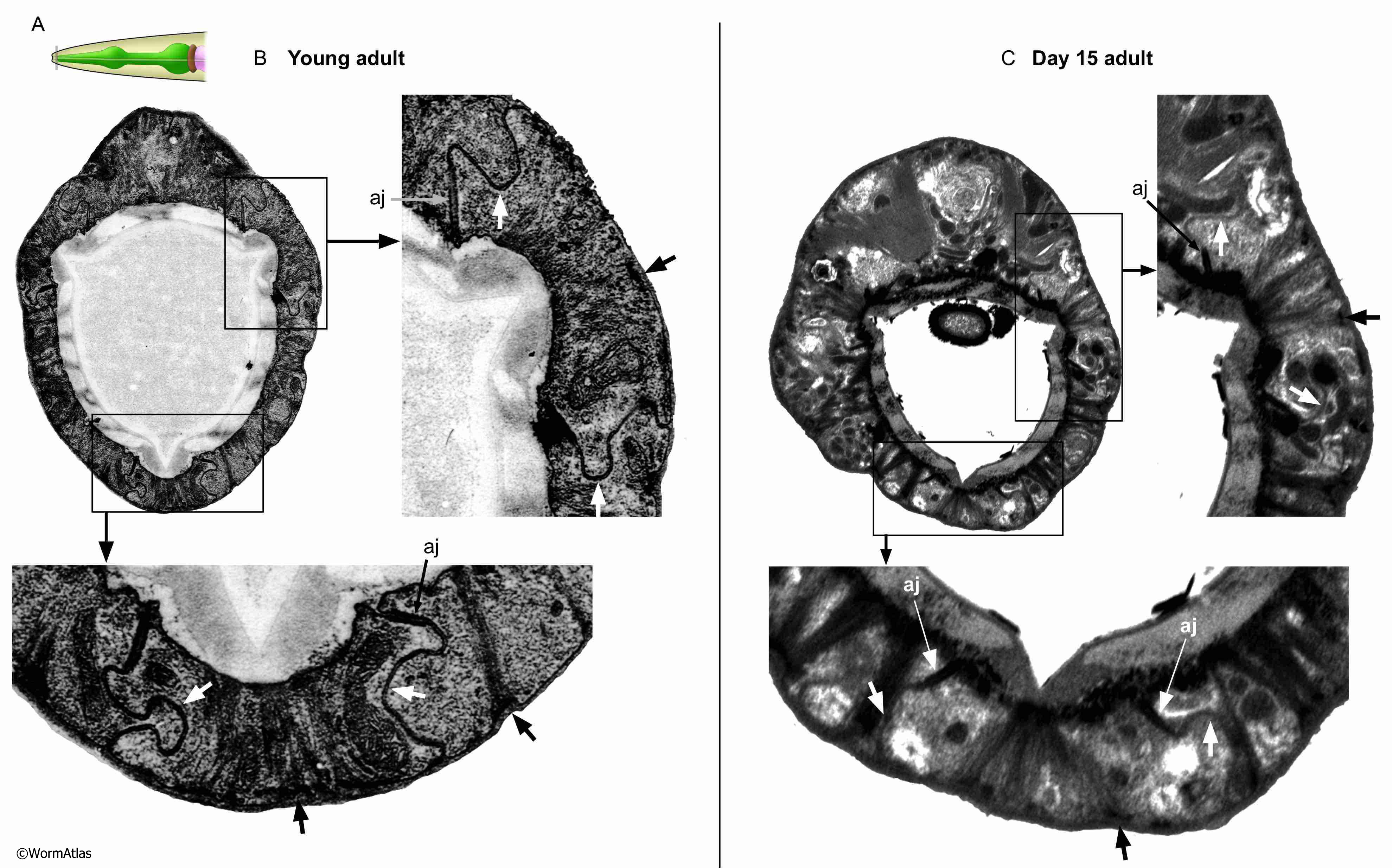
APhaFIG 5: Anterior pharynx epithelium in old adults.
Examination of cell junctions and features of anterior pharynx epithelial cells.
A. Illustration of C. elegans head showing approximate location of sections in B&C with grey bar. B. Young adult. C. 15-day-old adult.
B&C. Transverse section of the anterior pharyngeal epithelium. Rectangles indicate the expanded regions. Darkly-stained epithelial cell filaments persist in this old animal with at least partially intact hemidesmosome attachments to the basement membrane (black arrows). Lateral membranes between pharyngeal epithelial cells display large, smoothly curved, electron dense gap junctions in young adults (white arrows in B), but the same areas in older adults are less electron dense (white arrows in C) and less convincing to be intercellular junctions. Epithelial cells are connected to one another at the basement membrane by adherens junction (aj) which are also preserved in this older adult.
(Image source: B. N2T [N. Thomson] 089; C. N813 [D. Hall] G476.)
Click on picture for full resolution image.
|