|
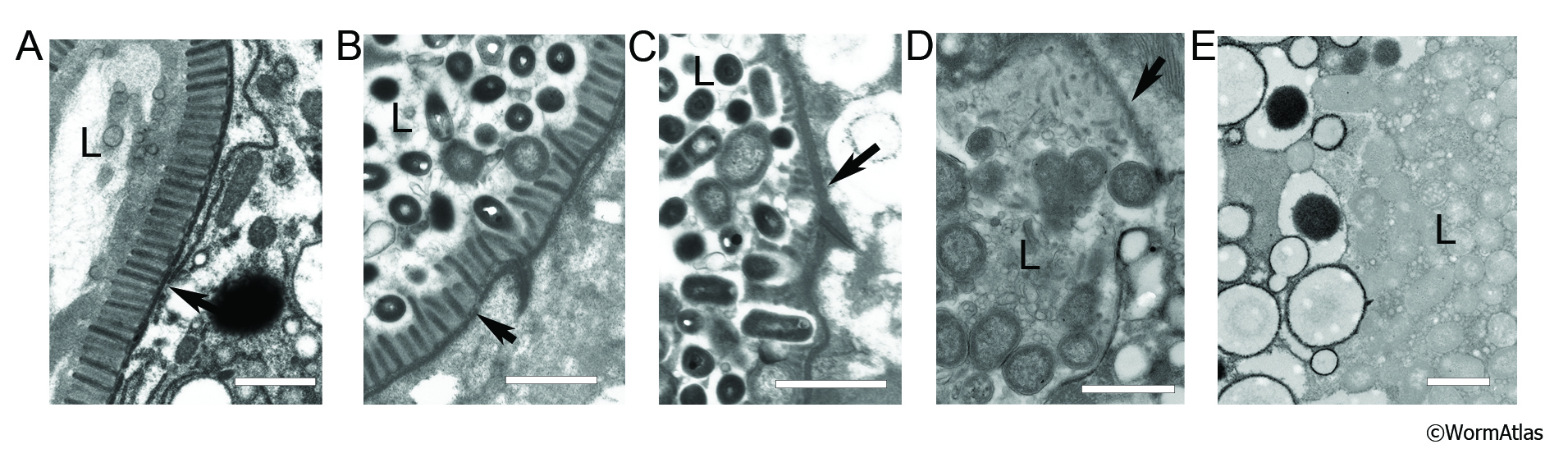
AIntFIG 6: Degradation of intestinal microvilli during aging.
Transverse TEM views displaying major defects in the adult day 7 intestinal microvilli in adulthood. All examples come from wild-type animals of the same age, with a wide variety of phenotypes. Progressive loss of several barriers to bacterial invasion of the cytoplasm are evident.
A. Healthy microvilli face a lumen filled mostly with soluble items or a few bacterial fragments. Arrow indicates a region where the terminal web may be separating from the base of the microvilli. (Image source: N826 [Hall] G4239). Bar, 1 µm.
B. Microvilli are no longer uniform in length, and intact bacteria can be seen in the lumen, some of which are attached to individual villi, possibly beginning to degrade them. Arrow indicates the terminal web. (Image source: N821 [Hall] 4872). Bar, 1 µm.
C. Most microvilli are gone, although the terminal web (arrow) remains thickened and electron dense. The lumen contains many intact bacteria. (Image source: [Hall] N821 4855). Bar, 1 µm.
D. The microvillar border and the terminal web (arrow) separating the lumen from the intestinal cytoplasm are less electron dense and possibly incomplete, allowing bacteria to invade the intestinal cell cytoplasm. (Image source: N831 [Hall] W006, class C). Bar, 1 µm.
E. The bacteria-filled lumen (L) (right) and the intestinal cytoplasm (left) seem to be in direct contact along an ill-defined interface, with no obvious structure to divide them. (Image source: N833 [Hall] W090 class B). Bar, 1 µm.
Click on picture for full resolution image.
|