|
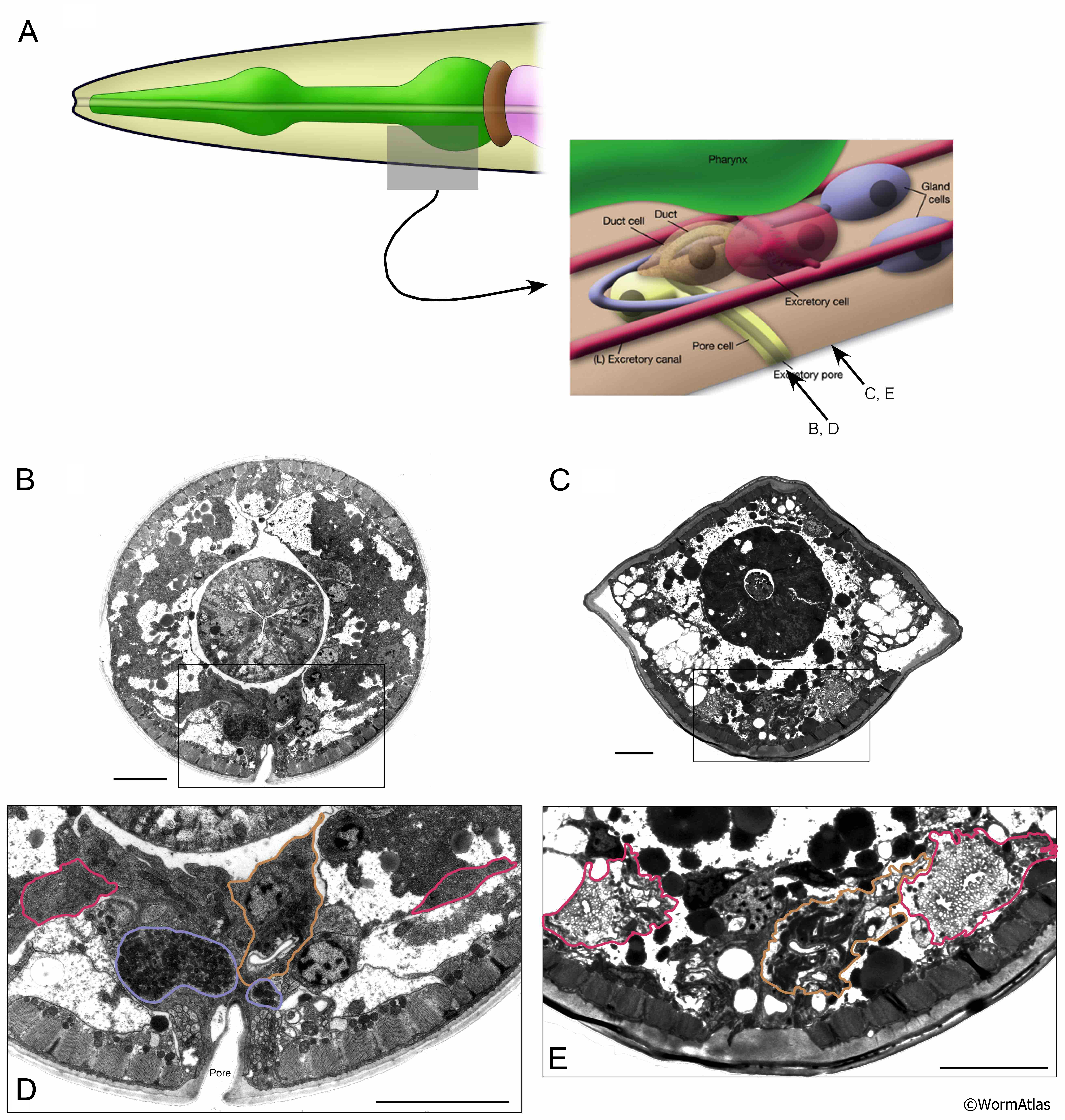
AExcFIG 7: Excretory sinus region in head of a day 7 adult.
A. Left, Diagram of the C. elegans head with region of the excretory sinus indicated by a grey box. Right, diagram of the excretory sinus region. Arrows indicate approximate positions of (B-E).
B,C. Transverse photomicrographs in excretory sinus region from young adult (B) and day 7 adult (C). Boxes outline the regions enlarged in (D&E).
D,E. Enlarged views of the excretory sinus regions from (B&C). Excretory structures are outlined. Excretory cells, pink; gland cells, purple; duct cell, brown. The excretory pore is visible in B&D.
C&E is positioned slightly posterior to the excretory pore. The excretory canals (pink) are enlarged and disorganized in the day 7 adult and appear to contain multiple lumens, or perhaps a single lumen that loops back and forth within the region. Canaliculi are maintained in the day 7 excretory canals, but are quite swollen. In the young adult, the gland cells (purple) contain abundant dark granule structures (D). In contrast, the gland cells were not apparent in the day 7 adult. The duct cell is well maintained in the day 7 adult (brown). The duct cell may have increased levels of cytoplasmic, darkly staining structures.
Bars: B,D,E: 5 microns; C: 10 microns. (Image source: B,D: N513A [D. Hall] 1463, C,E: N826 [D. Hall] 5767.)
Click on picture for full resolution image.
|