|
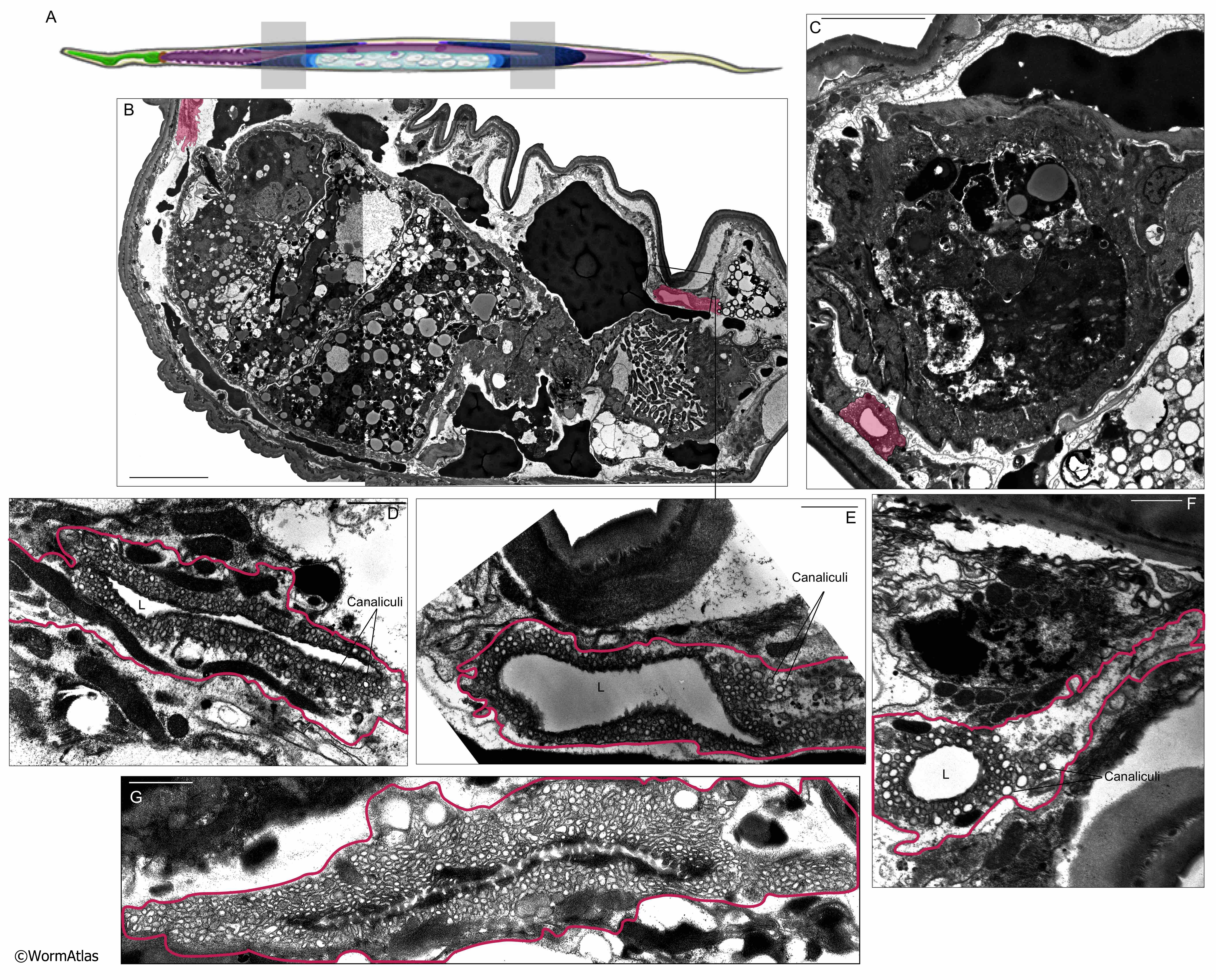
AExcFIG 6: Excretory canals in the midbody region of day 15 adults.
A. Diagram of the C. elegans body showing the two possible positions of panels B-F with grey boxes.
B. Oblique thin section of day-15 adult. Excretory canals are filled with pink. The apical border of one excretory canal is highly ruffled (left) but the other appears smoother (right). Both canals have enlarged lumens. Body cavity is filled by a tumorous germline mass. Bar, 10 microns, Image source: N803 [D. Hall] E763&764.
C. Excretory canal with an enlarged lumen of a day 15 adult. Bar, 5 microns. Image source: N803 [D. Hall] E805.
D-F. Excretory cells are outlined in pink. These cells all have enlarged lumens. Canaliculi are densest close to the apical border adjacent to the lumen. The basal borders are variably disorganized in the different animals, which are seen in oblique (D&E) or transverse (F) views.
G. Excretory cell showing disorganized basal border detached from the neighboring cells, longitudinal view. Bars, 1 micron. (Image source: D: N803 [D.Hall] E765, E: N803 [D. Hall] E766, F: N803 [D. Hall] E787, G: N811 [D. Hall] E297.)
Click on picture for full resolution image.
|