|
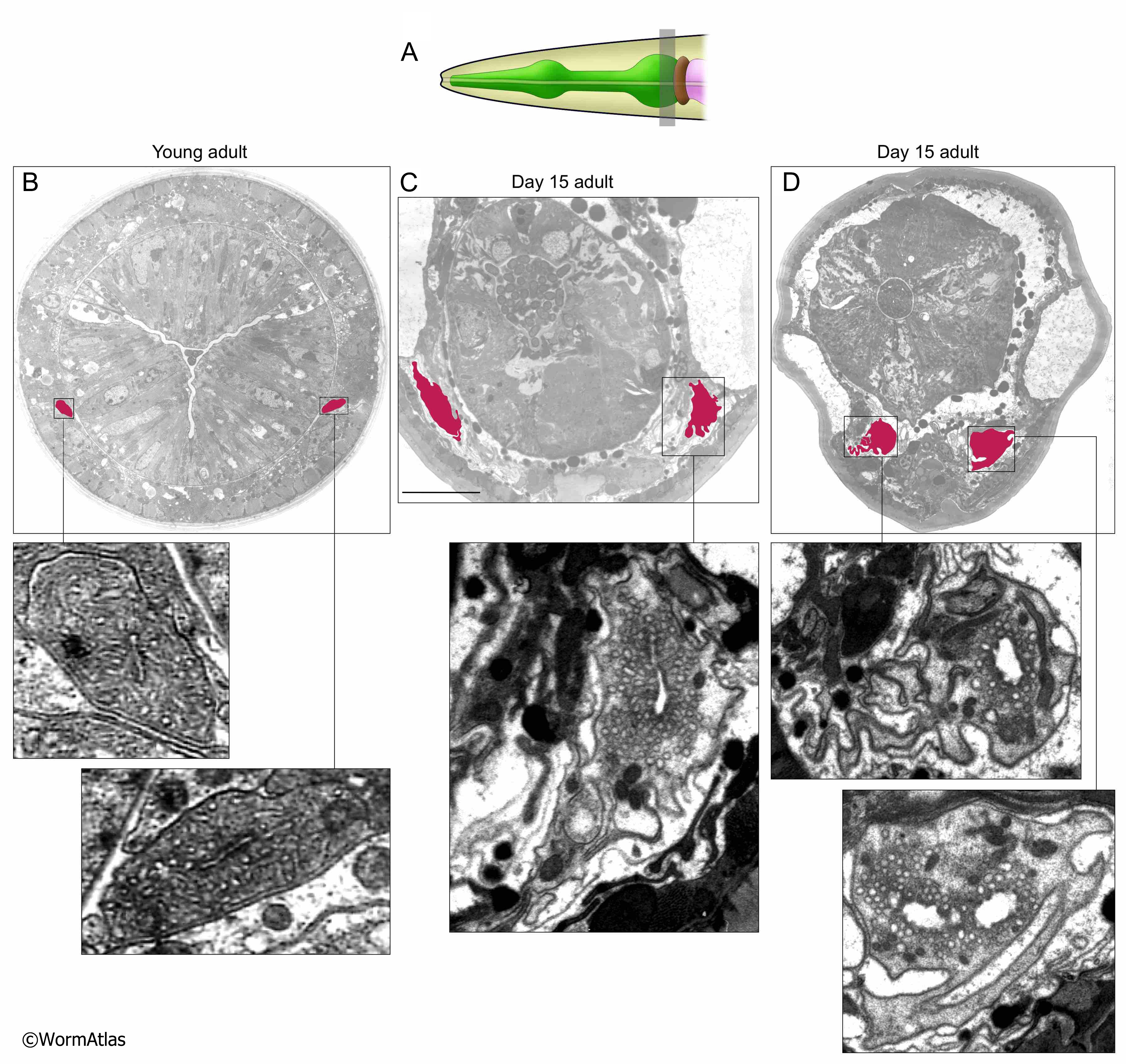
AExcFIG 4: Excretory cell structure near the posterior pharynx during aging.
A. Diagram of the C. elegans head showing the approximate positions of photomicrographs B,C&D by shaded box in the posterior portion of the pharynx adjacent to the pharygeal-intestinal valve.
B,C,D. Transverse sections from a young adult (B) and two day 15 adults (C&D). The bilateral excretory canals are colored in pink. The apical borders are smooth in the young adult, becoming disorganized and ruffled in both day-15 adults. Lower panels, closeups of individual excretory canals as shown. One excretory cell (D) contains two lumens, the other cells contain one lumen.
D. Both excretory canals appear to contain fewer canaliculi than the other animals.
C. Bar, 5 microns. (Image source: B: N513A [D. Hall] G534 M1 959, C: N829 [D. Hall] W001, D: N829 [D. Hall] R128).
Click on picture for full resolution image.
|