|
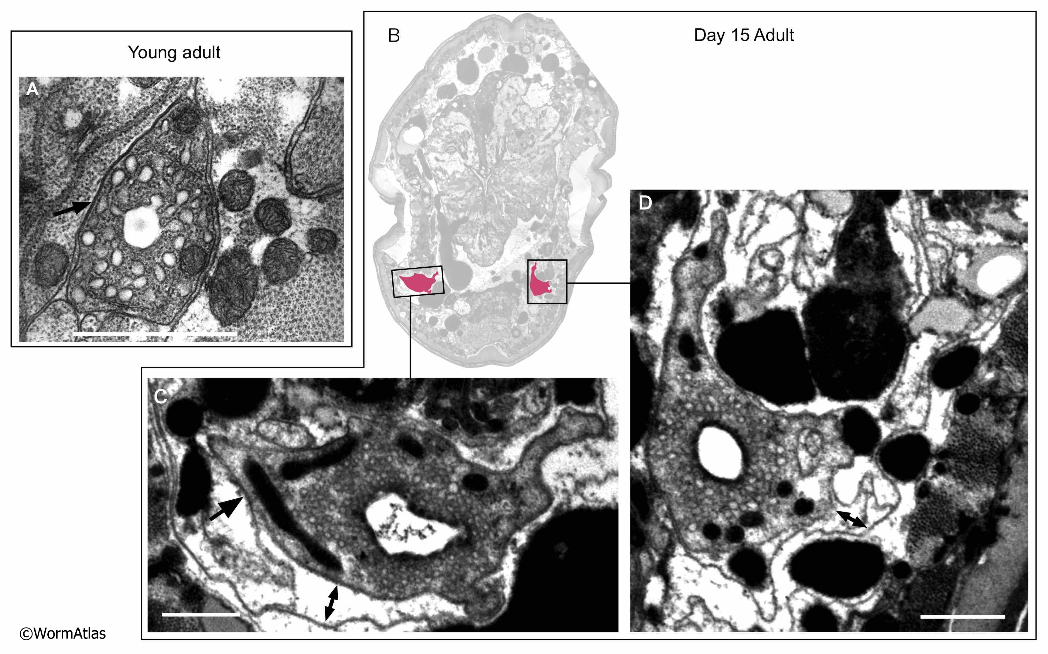
AExcFIG 3: Hypodermal-excretory cell attachments.
A. Excretory cell from a young adult showing extensive attachments with neighboring cells as darkened areas at the cell boundaries (arrow).
B. Low-magnification view of day-15 adult head. Positions of panels C&D are boxed, excretory cells shaded in pink.
C,D. Higher magnification view of excretory cells from (B). The hypodermal cells have lost much of their structure by this age. In some regions, attachments to the excretory cells may be maintained (arrows) but lost in other areas as the cell membranes appear to spread apart (double-headed arrows).
Bars, 1 micron. (Image source: A: N510A [D. Hall] V791, B. N813 [D. Hall] G548 & G549, C,D. N813 [D. Hall] G549.)
Click on picture for full resolution image.
|