Click pictures for higher resolution images Click pictures for higher resolution images
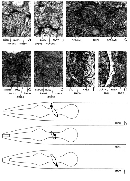
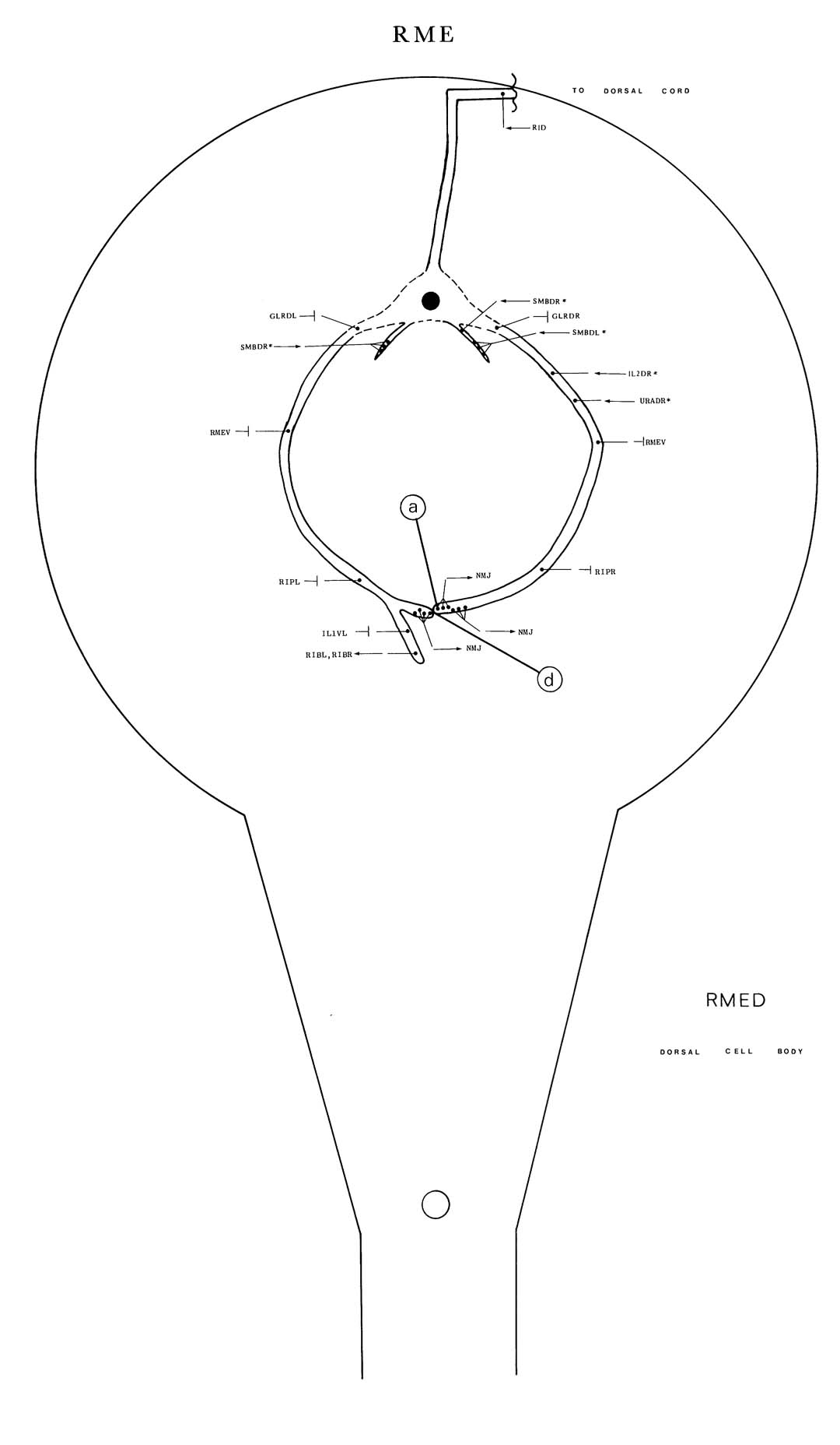
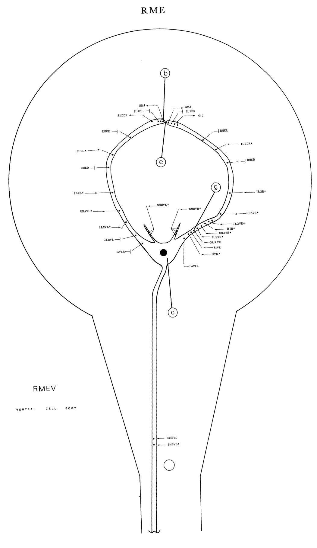
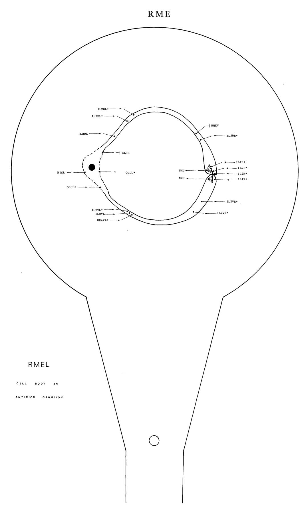
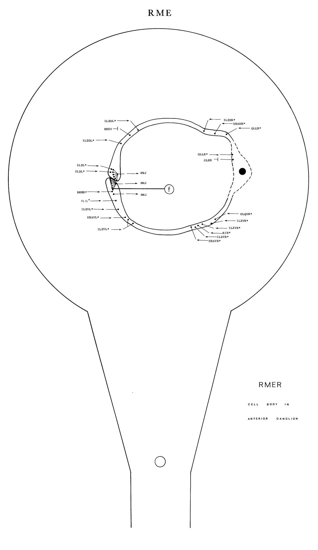
RME is a set of four motoneurons, with NMJs in the nerve ring, which innervate head muscles. The members of this class have an unusual fourfold symmetry; cell bodies are situated mid-dorsally (RMED), mid-ventrally (RMEV), left laterally (RMEL) and right laterally (RMER). Each cell has a twofold symmetry and sends out two processes, which run in opposite directions round the nerve ring near the anterior surface. These processes run alongside processes of adjacent RMEs and meet at the distal side of the nerve ring, forming NMJs at this point (d, e, f and figure 14). This meeting point is always located on the mid-line opposite to that on which the cell body is located. RMED/RMEV each send out an extra process; these run down the ventral and dorsal cords respectively, for about 150 um, and then peter out without making any significant synaptic connections along their length. RMED/RMEV also have two small dendrites, emanating from their cell bodies, which intercept the NMJs of SMB (*e). The cell bodies of all the RME neurons are closely apposed to the nerve ring and are ensheathed by the thin sheet-like processes of the CEPsh cells that surround the neuropile of the nerve ring (c). RMEL/RMER synapse exclusively onto muscle; RMED/RMEV synapse onto muscle (a, b) but probably also synapse onto adjacent SMD processes (d, e). RME receives synaptic input from IL2 (*c), SMB (*e) (RMED/RMEV only), OLL (RMEL/RMER only) and possibly URA (*c). There are gap junctions to itself, AVE, RIP and IL1D/IL1V (*g), and prominent gap junctions to the glial cells, GLR (g); RME is the only neuron class that makes contact with these cells (figure 15). Magnifications: (a, b) x 25500, (c-g) x 12750.

|
|