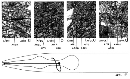
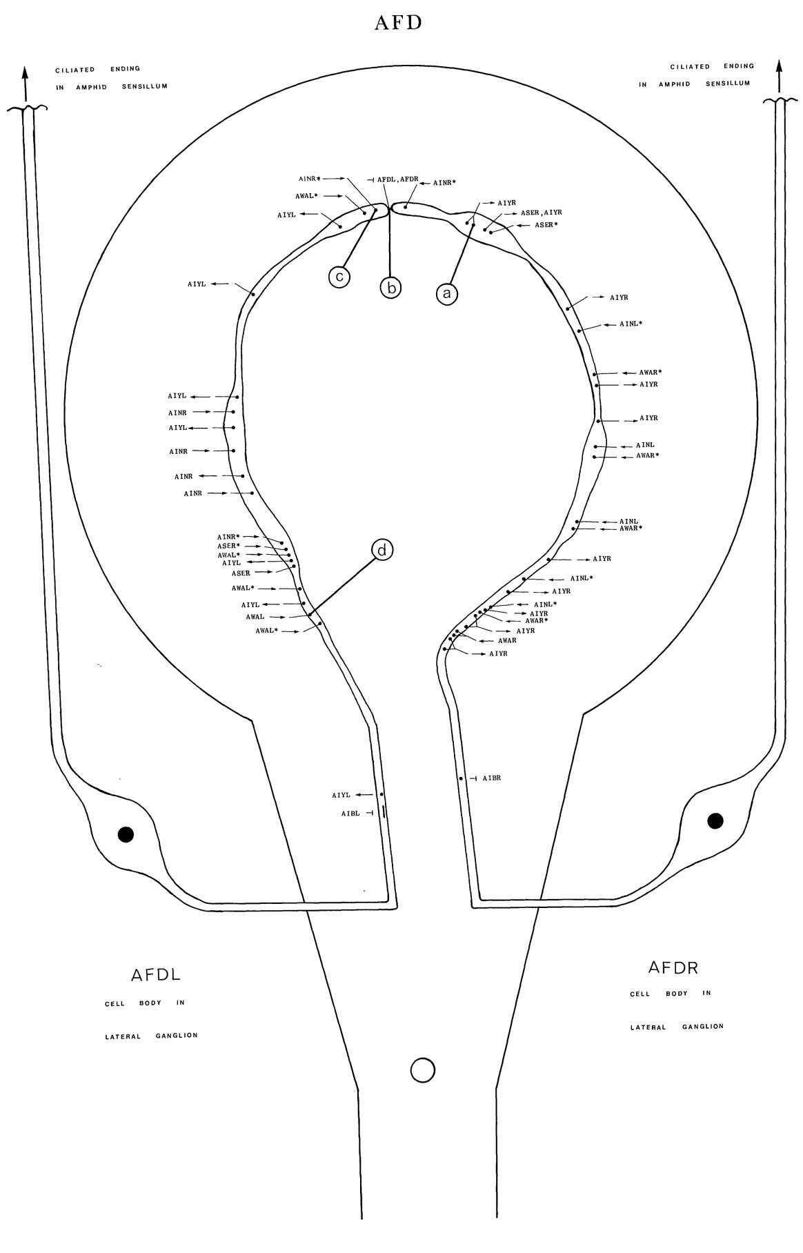
AFD is a set of two ciliated neurons that are part of the amphid sensillum. The endings of AFD have numerous villi, which poke into the amphid sheath cells (figure 1). The cell bodies are situated in the lateral ganglia; processes enter the ventral cord via the amphidial commissures and turn anteriorly to enter the nerve ring. They run round on the outside surface and the posterior face of the nerve ring in close association with the processes of AIY until they meet at the dorsal mid-line, where they terminate. There is a gap junction at the point of contact (b). The only synaptic output is to AIY (a); some dark-cored vesicles are seen in presynaptic terminals (a). There are synaptic inputs from AIN (c) and AWA (d) and small gap junctions to AIB in the ventral ganglion. Magnifications: (a) x 25500, (b-d) x 12750.


Click pictures for higher resolution images