|
Steps to Prepare Dissected Worms for SEM
by David Greenstein and Loren Hoffman
Protocol
- Select L4 hermaphrodites en masse.
- Grow overnight (12-16h) to adult stage.
- Transfer to unseeded plate to remove bacteria.
- Transfer to a watch glass with 0.5ml of 1X PBS.
- Wash worms with one change of 1XPBS.
- Dissect gonads in 1xPBS containing 0.05% tricane/0.005% tetramisole.
- Transfer to a 6 x 50mm borosilicate glass tube.
- Recover the gonads by centrifugation at 1000 rpm in a IEC clinical centrifuge for 2 min at room temperature.
- Digest the gonadal basal lamina by incubation with 20 - 40 units/ml of Type II collagenase in 1xPBS for 2-12 min at room temperature.
-
After digestion place gonads on ice and gently remove supernatant from gonads which had settled during incubation period.
- Because gonads are fragile until fixed add all solutions slowly. Wash gonads with cold 1x PBS, allowing the gonads to settle by gravity.
- Then fix the gonads with 3% glutaraldehyde in 0.1 M sodium phosphate buffer (pH 7.2) for 4 h on ice.
- After they are fixed, wash the gonads three times with cold 0.1 M sodium phosphate buffer (pH 7.0) containing 5% sucrose, incubating on ice for 15 min during each wash.
- After the final wash, pipet the gonads into a watch glass and hand select intact gonads under a dissecting scope.
- Postfix the gonads in 1% osmium tetroxide in 0.1 M sodium phosphate buffer (pH 7.2) for 1 h at room temperature.
- Rinse gonads in several changes of 0.1 M sodium phosphate buffer (pH 7.2).
- Dehydrate in an ethanol series (50, 70, 95, 100%).
- Dehydrate again in pure ethanol.
- Place gonads in the cooled chamber of a critical point drier with the samples being covered with ethanol. To prevent the gonads from being damage when the solvent enters or is vented from the chamber, place the gonads between several layers of filter paper cut with a standard hole punch.
- Transfer dried samples to aluminum stubs covered with double stick tape using a camel hair brush.
- Desiccate overnight and then coat with gold using a sputter coater.
Figures
 Click pictures for new window with figure and legend, click again for high resolution image Click pictures for new window with figure and legend, click again for high resolution image
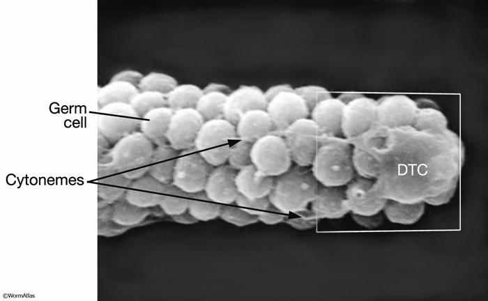
EMSEMDissectionFIG 1: Scanning electron micrograph of the DTC from dissected gonad. Lateral view. Distal-most end of an adult gonad (dissected away from the rest of the body). Long thin trailing processes of the DTC extend (leftward) across the unsheathed distal arm of the mitotic gonad. A bulge on the leading edge of the DTC (far right) may be the nucleus being pushed forward. (Image source: L. Hoffman and D. Greenstein, photo #003.)
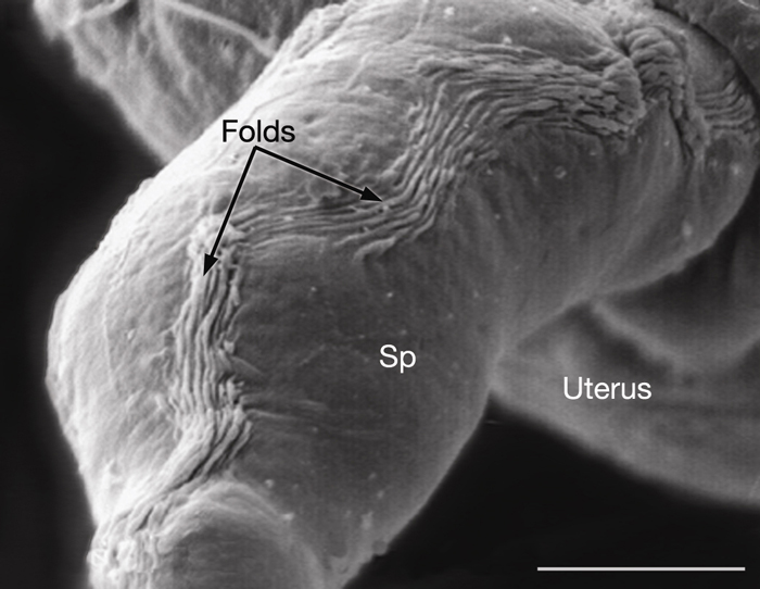
EMSEMDissectionFIG 2: Spermatheca viewed by SEM after dissection. Lateral view. Distal-most end of an adult gonad (dissected away from the rest of the body). Long thin trailing processes of the DTC extend (leftward) across the unsheathed distal arm of the mitotic gonad. A bulge on the leading edge of the DTC (far right) may be the nucleus being pushed forward. (Image source: L. Hoffman and D. Greenstein, photo #003.)
References
Hall, D.H., Winfrey, V.P., Blauer, G., Hoffman, L., Furuta, T., Rose, K.L., Hobert, O. and Greenstein, D. 1999. Ultrastructural features of the adult hermaphrodite gonad of C. elegans: Relations between the germ line and soma. Dev. Biol. 212: 101-123. Article
|