|
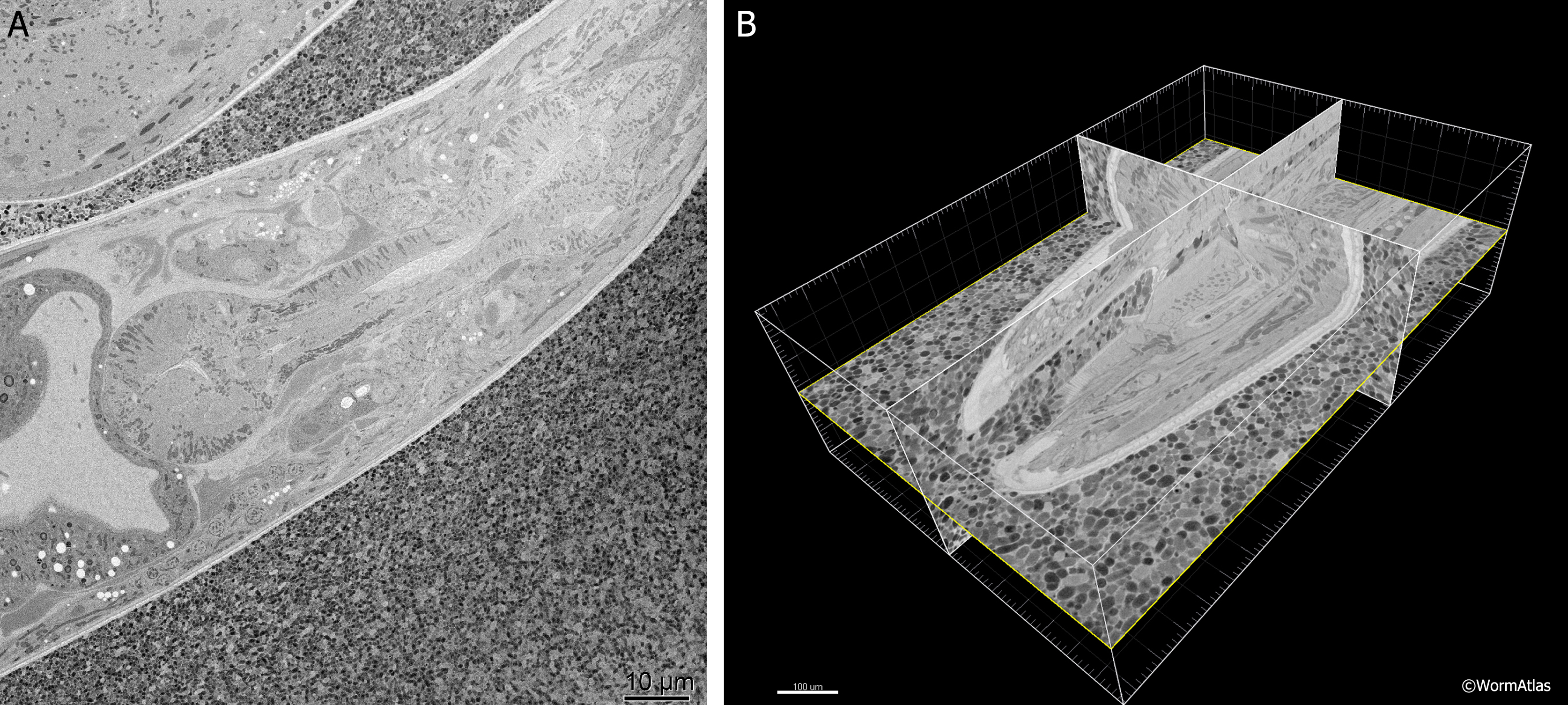
EMSerialblockFIG 1: Three-dimensional imaging from a ‘‘Denkotome.’’
A. Sample lengthwise view of an adult hermaphrodite head. HPF/FS sample. Thin sectioned inside a Gatan 3View. Image taken using back-scattered electrons to view the block face, not the thin section. Close-packed E. coli lie outside the nematode. Scale bar, 10 µm.
B. The same data are viewed from multiple angles by resampling the voxels in three dimensions. Here the original sections were collected in cross-section, but the 3D model is resampled in three different orthogonal planes. Bounding box indicates the region observed in serial images. Scale bar, 10 µm. Images provided courtesy of Joel Mancuso, Gatan Instruments.
Click on picture for full resolution image.
|