|
Pharynx Atlas:
The pictures that comprise the Pharynx Atlas are a focal series through the terminal bulb of the
pharynx. The worm is lying on its right side, with anterior towards the upper left and posterior towards the lower right. Dorsal is the upper right and ventral is the lower left. The first image is a
superficial cut through the very leftmost edge of the pharynx.
Subsequent images go consecutively deeper, until the last goes through
the very rightmost edge. The focal planes are not equally spaced. They
were chosen so that all the nuclei in the terminal bulb would be
clearly visible in at least one image.
Roll over images to identify individual pharyngeal cells. Click on cells to go to a new page with detailed information.
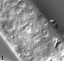
 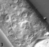 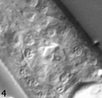 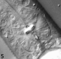 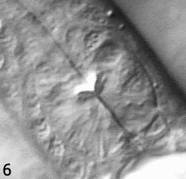 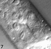 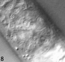
Image Section Key:
Image 1: 4 cells - M2L, mc3DL, pm6VL, pm7VL
Image 2: 9 cells - g1AL, g2L, I5, I6, M5, mc2DL, mc3V, pm5VL, pm8
Image 3: 9 cells - g1AL, g2L, I5, I6, M5, mc2DL, mc3V, pm5VL, pm8
Image 4: 10 cells - g1AL, g2L, I5, I6, M5, mc2DL, mc3V, pm5DL, pm5VL, pm8
Image 5: 3 cells - g2L, mc2V, pm5VL
Image 6: 5 cells - I4, mc2DR, pm5DL, pm6D, pm7D
Image 7: 7 cells - g1AR, M2R, pm5DR, pm5R, pm6D, pm6VR, pm7VR
Image 8: 4 cells - g1AR, g1P, M1, mc3DR
(Cells not visible in these planes: pm5L, pm5VR, g2R)
|
|
