Type: Motor neuron
In MoW: RME
Male Wiring Project:
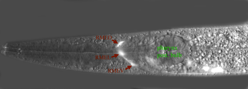
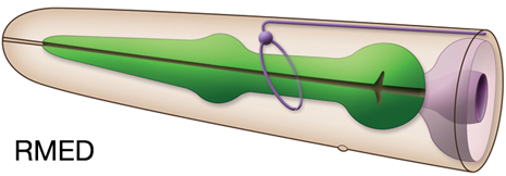
RMED,
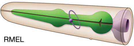
RMEL,
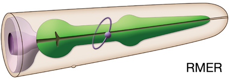
RMER,
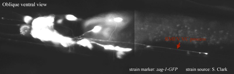
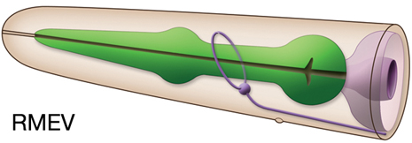
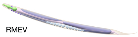
RMEV
In Wormbase: RME, RMED, RMEL, RMER, RMEV
Lineage: AB alapppaap, AB alaaaarlp,
AB alaaaarrp, AB plpappaaa
Location: Head
Description: Ring motor neurons which innervate head muscles with NMJs in the nerve ring
Neurotransmitter/ Neuropeptide:
- GABA
Innexin expression:
- UNC-7
- UNC-9
(Altun et al., 2009)
Receptor expression:
- ACR-14; nicotinic AChR non-alpha subunit
- AVR-15; glutamate-gated chloride channel subunit (only in RMED and RMEV)
- GLR-1 ; glutamate receptor subunit (only in RMEL and RMER)
- PDFR-1; pigment dispersing factor (PDF-1) receptor (only in RMED and RMEV)
-SER-2; a splice variant of the tyramine receptor
(Barrios et al., 2012; Fox et al., 2005; Tsalik et al., 2003; Dent et al., 1997; Hart et al., 1995)
Function: Suggested to be the pioneers of the nerve ring (Antebi et al., 1997)
|

|


 Click pictures for higher resolution images
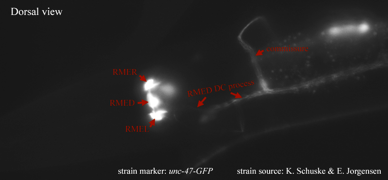

.jpg)
.jpg)
.jpg)