Type: Motor neuron
In MoW: RMD
Male Wiring Project:
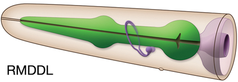
RMDDL,
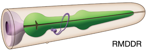
RMDDR,
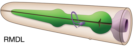
RMDL,
RMDR,
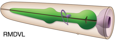
RMDVL,
RMDVR
In Wormbase: RMD, RMDDL, RMDDR, RMDL, RMDR, RMDVL, RMDVR
Lineage: AB alpapapaa, AB arappapaa, AB alpppapad, AB praaaapad, AB alppapaaa,
AB arapppaaa
Location: Head
Description: Ring motor neurons which innervate muscles in the head via NMJs in the nerve ring
Neurotransmitter/ Neuropeptide:
- Acetylcholine
(Although earlier reports suggested RMD might be glutamatergic, more recent studies showed it is not)
(Serrano-Saiz et al., 2013; Ohnishi et al., 2011; Loer, 2010; Duerr et al., 2008)
Innexin expression:
- UNC-7
(Altun et al., 2009)
Receptor expression:
- GLR-1; AMPA-type ionotropic glutamate receptor
(Maricq et al., 1995)
Function:
- IL1, OLQ, and RMD regulate spontaneous foraging movements;
in the presence of food, C. elegans makes rapid nose oscillations termed 'foraging' as they explore their environment.
Laser operated animals lacking IL1 and OLQ forage abnormally slowly and make exaggerated dorsal and ventral nose turns (Hart et al., 1995; Kaplan and Driscoll, 1997; Goodman, 2006.) See head withdrawal and foraging circuit.
- When worms are touched on the ventral or dorsal side of the nose during forward movement they respond with an aversive head movement away from the stimulus, termed the head-withdrawal reflex. This simple reflex is mediated by two classes of mechanosensory neurons (OLQ and IL1) and their synaptic targets, the RMD motor neurons. Killing any of these cells, alone or in combination, diminishes this reflex (Kaplan and Driscoll, 1997; Kindt et al., 2007.) TRPA-1 function in IL1 and OLQ and GLR-1 function in RMD neurons facilitate both foraging rate and head withdrawal (Kindt et al., 2007.)
|


|


