RIML, RIMR

Type: Motor neuron, interneuron
In MoW: RIM
Male Wiring Project:
RIML, RIMR
In Wormbase: RIM, RIML, RIMR
Lineage: AB plppaapap, AB prppaapap
Location: Lateral ganglia of head
Description: RIM neurons send processes into the nerve ring via amphid commissures. They innervate muscles in the head via NMJs in the nerve ring
Neurotransmitter/ Neuropeptide:
- Glutamate
- Tyramine
(Loer, 2010; Alkema et al., 2005)
Innexin expression:
- INX-4
(Altun et al., 2009)
Function: RIM, RIA and RIB are classified as the "second layer" interneurons in the process of integration of information from the outside world and the inner state of the animal, which then leads to a behavioral response.


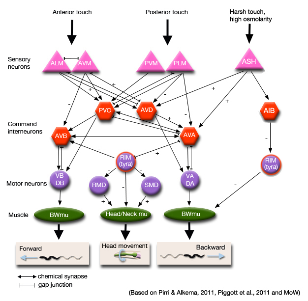
- Function in the locomotion circuit; RIM neurons modulate reversals by two opposing roles. Photostimulation of RIM induces the worm to reverse and this action requires AVA/AVD/AVE neurons (Guo et al., 2009). RIM and AVA activity are correlated through gap juntions, while RIM inhibits AVB (Alkema et al., 2005, Pirri et al., 2009). Anterior touch activates AVA, which, in turn, activates RIM. Tyramine release from the RIM neurons activates the tyramine-gated chloride channel LGC-55 on the AVB forward locomotion command neurons, neurons of the head movement circuit, RMD and SMD, and the neck muscles. Activation of LGC-55 causes hyperpolarization of the neck muscles and the AVB neurons inducing the suppression of head movements and sustained backward locomotion (ie reversal) in response to anterior touch (Pirri and Alkema, 2011. It was also shown, however, that optogenetic ablation of RIM increases reversal frequency, which suggests RIM inhibits initiation of reversals during locomotion (Piggott et al,, 2011; Husson et al., 2013). Suppression of RIM activity can trigger reversals independently of the command interneurons for backward locomotion, i.e. AVA/AVD/AVE, through a disinhibition circuit that involves AIB upstream of RIM. This is considered a parallel pathway for reversal initiation.


Click image for closeup view Click pictures for higher resolution images


Click here for larger version
RIML (AB plppaapap) development in the embryo.
Dorsal view. Bottom is left side of the embryo. Spheres indicate individual nuclei. Black sphere: ancestors of RIML; dark grey spheres: apoptotic cells; other cells follow the WA color code (after they acquire specific cell or tissue identities). 0 min is fertilization. Click on the movie for higher resolution rendition (by A. Santella & Z. Bao).


Click here for larger version
RIMR (AB prppaapap) development in the embryo.
Dorsal view. Bottom is left side of the embryo. Spheres indicate individual nuclei. Black sphere: ancestors of RIMR; dark grey spheres: apoptotic cells; other cells follow the WA color code (after they acquire specific cell or tissue identities). 0 min is fertilization. Click on the movie for higher resolution rendition (by A. Santella & Z. Bao).


Last revision: July 8, 2014
image