|
Click here for larger version
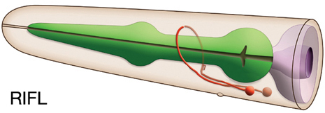
RIFL (AB plppapaaap) development in the embryo. Dorsal view. Bottom is left side of the embryo. Spheres indicate individual nuclei. Black sphere: ancestors of RIFL (since last RIFL ancestor has not yet gone through its final division, the black sphere seen at the end of this movie is still AB plppapaaa); dark grey spheres: apoptotic cells; other cells follow the WA color code (after they acquire specific cell or tissue identities). 0 min is fertilization. Click on the movie for higher resolution rendition (by A. Santella & Z. Bao). |
Click here for larger version
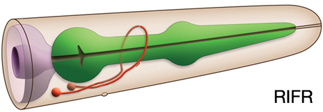
RIFR (AB prppapaaap) development in the embryo. Dorsal view. Bottom is left side of the embryo. Spheres indicate individual nuclei. Black sphere: ancestors of RIFR (since last RIFR ancestor has not yet gone through its final division, the black sphere seen at the end of this movie is still AB prppapaaa); dark grey spheres: apoptotic cells; other cells follow the WA color code (after they acquire specific cell or tissue identities). 0 min is fertilization. Click on the movie for higher resolution rendition (by A. Santella & Z. Bao). |