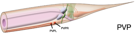
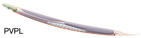
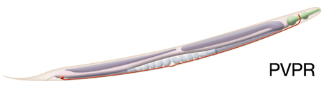
PVPL (called PVU in male), PVPR (called PVS in male)

Type: Interneuron
In MoW: PVP
Male Wiring Project:
PVU, PVS
In Wormbase: PVP, PVPL, PVPR
Lineage: AB plppppaaa, AB prppppaaa
Location: Pre-anal ganglion
Description: Interneuron. Differences in connectivity observed between hermaphrodites and males. In hermaphrodites PVP neurites were observed to make branches around the vulva suggesting they may form synapses with egg-laying muscles (Hobert and Hall, 1999). The PVPL/PVPR processes cross over in the pre-anal ganglion and then grow forward on the opposite side of the cord to the nerve ring. The PVPL process invariably crosses in front of the PVPR process. PVPL and PVPR somas are nearly opposite one another in the embryo and early larval stages. However, in the adult, this symmetry and order is lost and cells are situated almost linearly within PAG (Durbin, 1987).


Neurotransmitter/ Neuropeptide:
- Acetylcholine
(Duerr et al, 2008)
Innexin expression:
- INX-7
- INX-10
- UNC-7
- UNC-9
(Altun et al., 2009)
Receptor expression:
Function:
- PVPR pioneers the left tract of the ventral cord and this function appears to be a joint action of PVPR and PVQL processes; PVQ axons pioneer the lumber commissures and continue to travel alongside the PVP processes in the VNC (Durbin, 1987; Wadsworth and Hedgecock, 1996; Wadsworth et al., 1996). When PVPR precursor is ablated in the embryo, PVQL and AVKR growth cones migrate towards the right side. (Durbin, 1987; Antebi et al., 1997)


Click image for closeup view Click pictures for higher resolution images
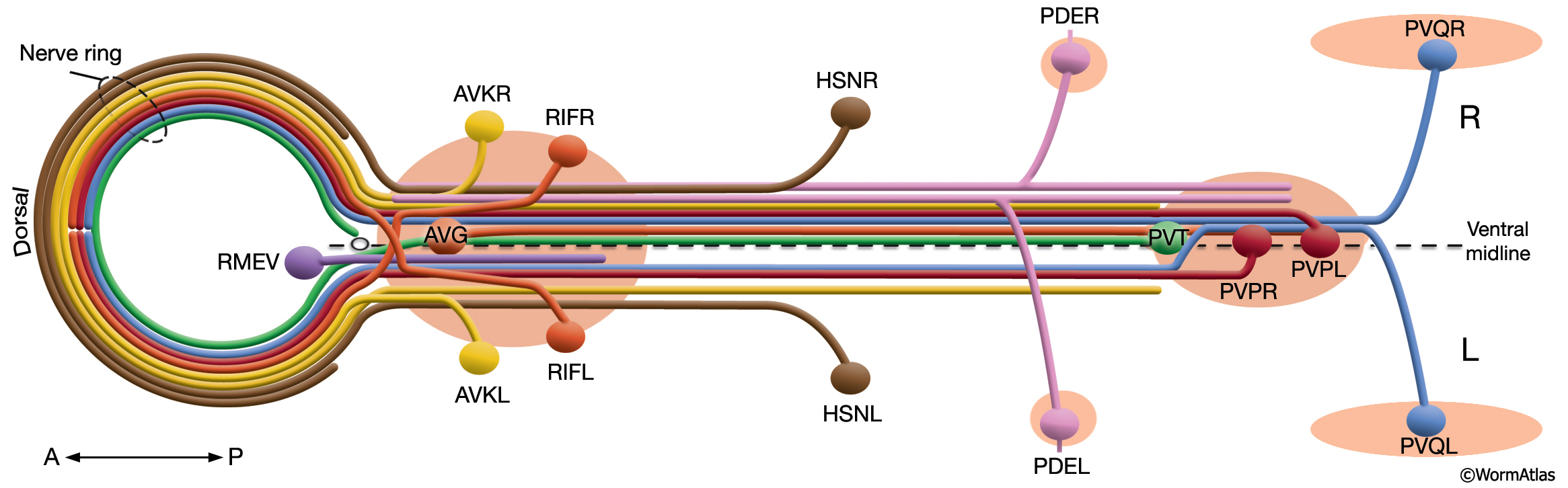
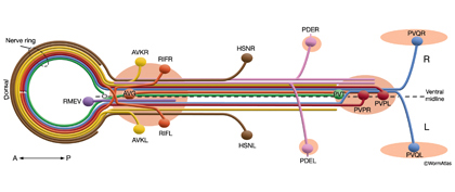
Schematic view of neurons that pioneer the left and right fascicles of the VNC. The body posterior to the NR is shown as if opened along the dorsal midline in cylindrical projection while the nerve ring is flattened toward the anterior. (Circle) Excretory pore at the anterior of the ventral midline; (light red ovals) ganglia. For clarity, neurons are given individual colors that are different from the color code used throughout this Atlas. (Based on White et al., 1986; Wadsworth et al., 1996.)


Click here for larger version
PVPL (AB plppppaaa) development in the embryo.
Dorsal view. Bottom is left side of the embryo. Spheres indicate individual nuclei. Black sphere: ancestors of PVPL; dark grey spheres: apoptotic cells; other cells follow the WA color code (after they acquire specific cell or tissue identities). 0 min is fertilization. Click on the movie for higher resolution rendition (by A. Santella & Z. Bao).


Click here for larger version
PVPR (AB prppppaaa) development in the embryo.
Dorsal view. Bottom is left side of the embryo. Spheres indicate individual nuclei. Black sphere: ancestors of PVPR; dark grey spheres: apoptotic cells; other cells follow the WA color code (after they acquire specific cell or tissue identities). 0 min is fertilization. Click on the movie for higher resolution rendition (by A. Santella & Z. Bao).


Last revision: March 11, 2015
image