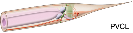
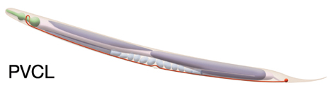
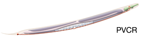
Type: Ventral cord interneuron
In MoW: PVC
Male Wiring Project:
PVCL,
PVCR
In Wormbase: PVC, PVCL, PVCR
Lineage: AB plpppaapaa, AB prpppaapaa
Location: Tail; lumbar ganglion
Description: PVC processes enter the nerve cord via the lumbar commissures and run on the right side of the hypodermal ridge in the nerve cord. PVCL process decussates posterior to the excretory pore and enters the nerve ring on the left side, while PVCR process enters the ring on the right. They run near the middle of the neuropil to make a full turn within the nerve ring before ending ventrally on the opposite side.
Neurotransmitter/ Neuropeptide:
- Acetylcholine
(Pereira et al., 2015; Duerr et al, 2008)
Innexin expression:
- INX-7
- INX-13
- INX-19
- UNC-9
(Schumacher et al., 2012; Altun et al., 2009; Chuang et al, 2007)
Receptor expression:
- PDFR-1; pigment dispersing factor (PDF-1) receptor
(Barrios et al., 2012)
Function: Command interneuron
- Functions as command interneuron for forward locomotion; drives forward movement of the animal along with AVB
- Modulates response to harsh touch to tail. Harsh tail touch induces Ca2+ increases in PVC, which are higher than those evoked by gentle touch, and a harsh touch defect is seen in the absence of PVC neurons. PVC may be sensing the stimulus directly or indirectly through other neurons (Li et al., 2011)
|
|


