- MEC-6; part of the degenerin/epithelial Na+channel complex - interacts physically with the MEC-4 degenerin ion channel
- TRPA-1; transient receptor potential (TRPA) ion channel protein (Wormbase; Kindt et al., 2007; Chelur et al., 2002; Treinin and Chalfie, 1995) Function:
- When worms are touched on either the dorsal or ventral sides of their nose with an eyelash, they interrupt the normal pattern of foraging and undergo an aversive head-withdrawal reflex. This simple reflex is mediated by two classes of mechanosensory neurons (OLQ and IL1) and their synaptic targets, the RMD motor neurons. Killing any of these cells, alone or in combination, diminishes the head withdrawal reflex. IL1, OLQ, and RMD also regulate spontaneous foraging movements. Laser operated animals lacking IL1 and OLQ forage abnormally slowly and make exaggerated dorsal and ventral nose turns (Goodman, 2006; Kaplan and Driscoll, 1997; Hart et al., 1995). See head withdrawal and foraging circuit.
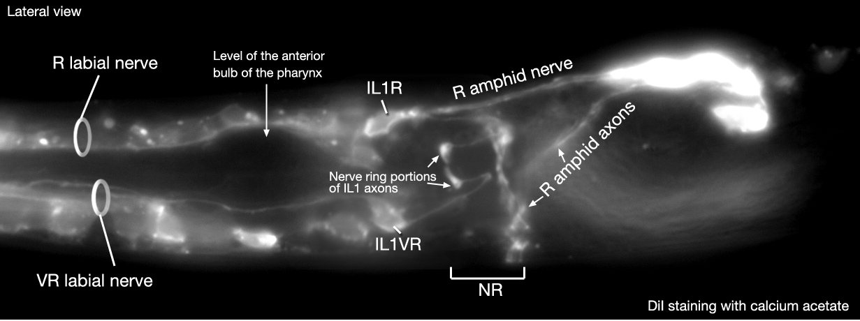
Click pictures for higher resolution images and to play movie
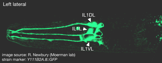
(3-D reconstruction of IL1 cells was created from confocal images of a strain expressing the GFP marker linked to the promoter for Y111B2A.8 using Zeiss LSM 5 Pascal software v. 3.2 by Newbury and Moerman lab)Embryonic development of IL1 neurons
Click here for larger version
IL1L (AB alapaappaa) development in the embryo. Dorsal view. Bottom is left side of the embryo. Spheres indicate individual nuclei. Black sphere: ancestors of IL1L (since last IL1L ancestor has not yet gone through its final division, the black sphere seen at the end of this movie is still AB alapaappa); dark grey spheres: apoptotic cells; other cells follow the WA color code (after they acquire specific cell or tissue identities). 0 min is fertilization. Click on the movie for higher resolution rendition (by A. Santella & Z. Bao).
Click here for larger version
IL1R (AB alaappppaa) development in the embryo. Dorsal view. Bottom is left side of the embryo. Spheres indicate individual nuclei. Black sphere: ancestors of IL1R (since last IL1R ancestor has not yet gone through its final division, the black sphere seen at the end of this movie is still AB alaappppa); dark grey spheres: apoptotic cells; other cells follow the WA color code (after they acquire specific cell or tissue identities). 0 min is fertilization. Click on the movie for higher resolution rendition (by A. Santella & Z. Bao).
Click here for larger version
IL1VL (AB alppapppaa) development in the embryo. Dorsal view. Bottom is left side of the embryo. Spheres indicate individual nuclei. Black sphere: ancestors of IL1VL (since last IL1VL ancestor has not yet gone through its final division, the black sphere seen at the end of this movie is still AB alppapppa); dark grey spheres: apoptotic cells; other cells follow the WA color code (after they acquire specific cell or tissue identities). 0 min is fertilization. Click on the movie for higher resolution rendition (by A. Santella & Z. Bao).
Click here for larger version
IL1VR (AB arapppppaa) development in the embryo. Dorsal view. Bottom is left side of the embryo. Spheres indicate individual nuclei. Black sphere: ancestors of IL1VR (since last IL1VR ancestor has not yet gone through its final division, the black sphere seen at the end of this movie is still AB arapppppa); dark grey spheres: apoptotic cells; other cells follow the WA color code (after they acquire specific cell or tissue identities). 0 min is fertilization. Click on the movie for higher resolution rendition (by A. Santella & Z. Bao).
Click here for larger version
IL1DL (AB alapappaaa) development in the embryo. Dorsal view. Bottom is left side of the embryo. Spheres indicate individual nuclei. Black sphere: ancestors of IL1DL (since last IL1DL ancestor has not yet gone through its final division, the black sphere seen at the end of this movie is still AB alapappaa); dark grey spheres: apoptotic cells; other cells follow the WA color code (after they acquire specific cell or tissue identities). 0 min is fertilization. Click on the movie for higher resolution rendition (by A. Santella & Z. Bao).
Click here for larger version
IL1DR (AB alappppaaa) development in the embryo. Dorsal view. Bottom is left side of the embryo. Spheres indicate individual nuclei. Black sphere: ancestors of IL1DR (since last IL1DR ancestor has not yet gone through its final division, the black sphere seen at the end of this movie is still AB alappppaa); dark grey spheres: apoptotic cells; other cells follow the WA color code (after they acquire specific cell or tissue identities). 0 min is fertilization. Click on the movie for higher resolution rendition (by A. Santella & Z. Bao).
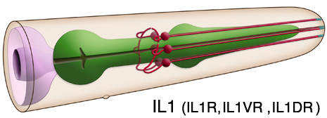
IL1 Sensory Endings
Click here for larger version
3D reconstruction of the anterior sensory endings (cilia and dendrites) from high resolution serial section transmission electron micrographs (ssTEMs). Bar 1 μm. Color code for the sensory endings is shown on the right-colors do not follow the WA color code. To expand, double click on the video, to return to original size, click "esc" (Doroquez et al., 2014)
Click here for larger version
3D reconstruction of all amphid neuron cilia and associated socket and sheath cell processes. Modeled from serial section transmission electron micrographs (ssTEMs). Bar 1 μm. Color code for the sensory endings is shown on the left-colors do not follow the WA color code. To expand, double click on the video, to return to original size, click "esc" (Doroquez et al., 2014)
Click here for larger version
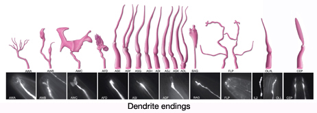
3D reconstruction of IL1, IL2, OLL, OLQ and CEP sensory endings and associated glia. Colors do not follow the WA color code (purple; transition zone (TZ), lavender; CEP, blue; OLL and OLQ, beige; IL1, green; IL2, light pink; ILsh, dark pink; ILso, dark orange; OLso, light orange; OLsh, dark green; CEPso, light green; CEPsh).To expand, double click on the video, to return to original size, click "esc" (Doroquez et al., 2014)


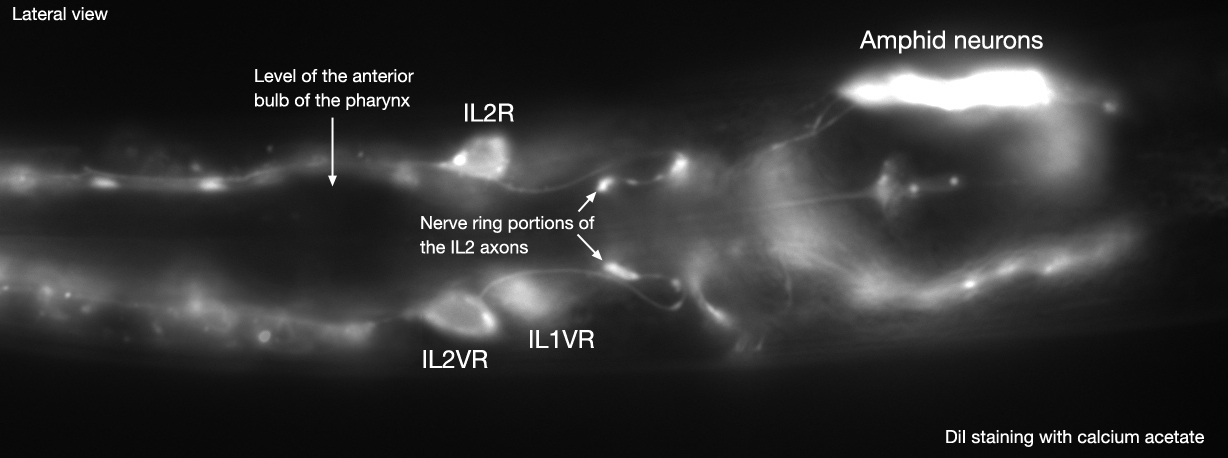
 Click pictures for higher resolution images and to play movie