Type:Sensory neuron (polymodal nociceptive for mechanosensation and thermosensation). Connectivity:
In MoW: FLP
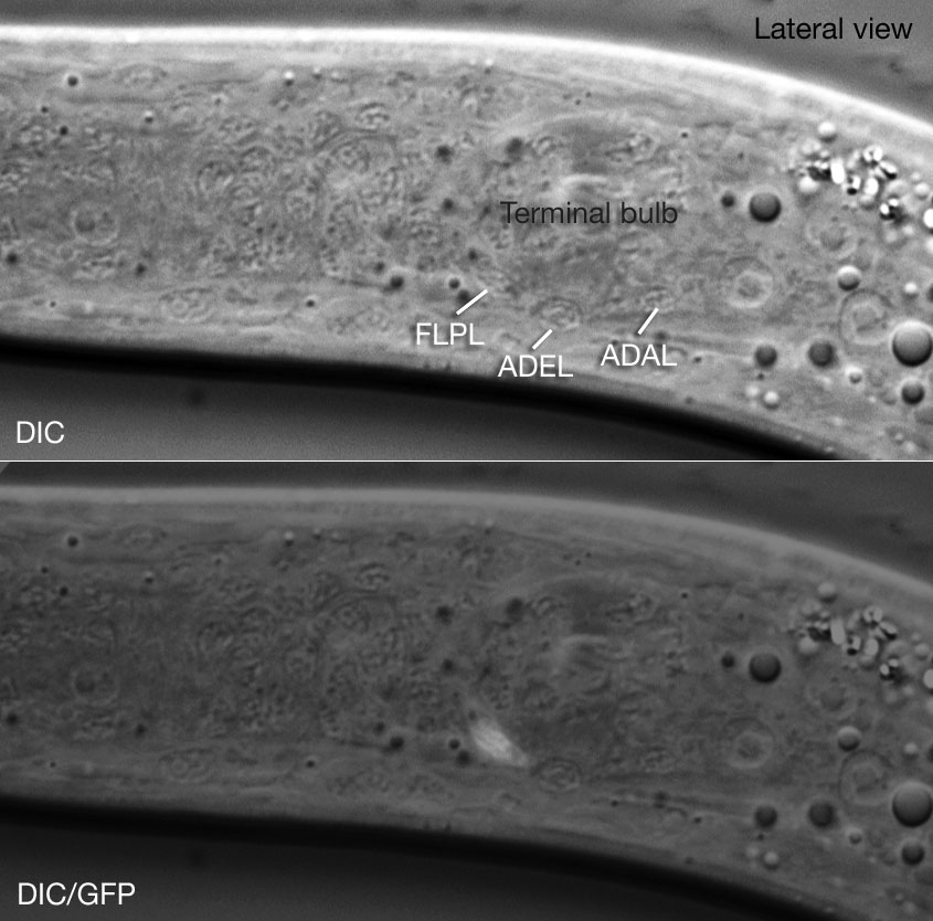
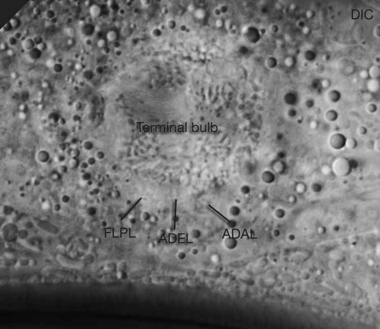
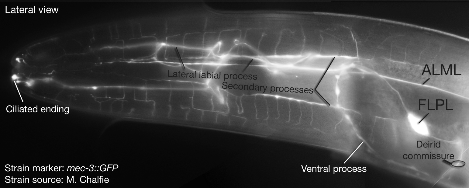
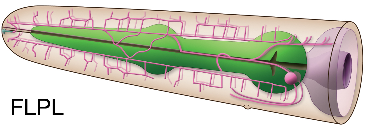
In Nemanode: FLP In Wormbase: FLP, FLPL, FLPR Lineage:AB plapaaapad, AB prapaaapad Location:Head Description: Name originally derived from "FLAP". Not part of a sensillum. FLP neurons are multidendritic with an arborization structure very similar to PVD neurons (Hall & Treinin, 2011; Hall & Altun, 2008). Topologically they cover head and neck, while PVD neurons cover the rest of the body (Hall & Altun, 2008). Their primary dendrites have ciliated endings in head which are not open to outside. They wrap around a branch of the lateral ILso cell just below the region where the socket cell surrounds the IL neurons. Neurotransmitter/ Neuropeptide:
- Glutamate
- FLP-4; FMRFamide-like neuropeptide
- FLP-21; FMRFamide-like neuropeptide (Loer and Rand, 2022; Li and Kim, 2008;Lee et al., 1999) Innexin expression:
- CHE-7
- INX-1a
- INX-1b
- INX-2
- INX-7
- INX-10a (only in dauer)
- INX-14
- INX-18a
- UNC-7
- UNC-9 (Altun et al., 2009; Bhattacharya et al., 2019) Receptor expression:
- DEG-3; alpha subunit of nicotinic acetylcholine receptor
- DEL-1; mechanically gated ion channel subunit; amiloride-sensitive Na+ channel (ASC) protein (DEG/ENaC subunit).
- DES-2; alpha subunit of nicotinic acetylcholine receptor
- GLR-4 ; putative non-NMDA ionotropic glutamate receptor subunit
- MEC-10; mechanically gated ion channel subunit; amiloride-sensitive Na+ channel (ASC) protein (DEG/ENaC subunit). Part of a mechanosensory transduction channel that senses low-threshold stimuli (gentle body touch)
- OSM-9; TRPV (transient receptor potential channel, vanilloid subfamily; mammalian capsaicin receptor-like channel)-cation selective. Among "thermoTRPs" (TRPA, TRPM, TRPV)
- PDFR-1; G-protein coupled class B subfamily B1 hormone receptor. Orthologous to drosophila pigment dispensing factor receptors and human CALCR
- UNC-8; mechanically gated ion channel subunit; amiloride-sensitive Na+ channel (ASC) protein (DEG/ENaC subunit) (Wormbase; Altun, 2011; Brockie et al., 2001; Treinin. et al., 1998; Colbert et al., 1997; Tavernarakis et al., 1997; Treinin and Chalfie, 1995; Huang and Chalfie, 1994). Function:
- Harsh nose touch response; nose-on collision with an object such as an eyelash initiates backward movement of C. elegans. Three classes of mechanosensory neurons (ASH, FLP, and OLQ) act in parallel to mediate this response. Each sensory neuron class accounts for a fraction of the normal response, as follows: ASH, 45%; FLP, 29%; and OLQ, 5%. The remaining responses (~10%) are mediated by the ALM and AVM neurons, which sense anterior body touch (Goodman, 2006; Kaplan and Driscoll, 1997)
- FLP neurons also respond to gentle nose touch and activate an escape behavior (Chatzigeorgiou and Schafer, 2011). OLQ and CEP neurons indirectly facilitate gentle nose touch responses in the FLP nociceptors via the RIH interneuron which acts as the integrating neuron of this circuit hub (Chatzigeorgiou and Schafer, 2011).
- AFD and FLP neurons in the head, and PHC neurons in the tail sense noxious temperatures (thermonociception) (~35-38oC) inducing a reflex-like escape reaction (temperature avoidance response). This avoidance response requires the cell autonomous function of cGMP dependent cyclic nucleotide-gated channels (TAX-2, TAX-4) in AFD, and the heat- and capsaicin-sensitive Transient Receptor Potential Vanilloid (TRPV) channels (OCR-2/OSM-9) in the FLP and PHC neurons (Liu et al., 2012). Reporters:
-At CGC: CF702muIs32 [mec-7p::GFP + lin-15(+)]. Also expressed in touch receptor neurons.
- Other:
Click pictures for higher resolution images
Click here for larger version
FLPL (AB plapaaapad) development in the embryo. Dorsal view. Bottom is left side of the embryo. Spheres indicate individual nuclei. Black sphere: ancestors of FLPL (since last FLPL ancestor has not yet gone through its final division, the black sphere seen at the end of this movie is still AB plapaaapa); dark grey spheres: apoptotic cells; other cells follow the WA color code (after they acquire specific cell or tissue identities). 0 min is fertilization. Click on the movie for higher resolution rendition (by A. Santella & Z. Bao).
Click here for larger version
FLPR (AB prapaaapad) development in the embryo. Dorsal view. Bottom is left side of the embryo. Spheres indicate individual nuclei. Black sphere: ancestors of FLPR (since last FLPR ancestor has not yet gone through its final division, the black sphere seen at the end of this movie is still AB prapaaapa); dark grey spheres: apoptotic cells; other cells follow the WA color code (after they acquire specific cell or tissue identities). 0 min is fertilization. Click on the movie for higher resolution rendition (by A. Santella & Z. Bao).
Click here for larger version
3D reconstruction of the anterior sensory endings (cilia and dendrites) from high resolution serial section transmission electron micrographs. Bar 1 μm. Color code for the sensory endings is shown on the right-colors do not follow the WA color code. To expand, double click on the video, to return to original size, click "esc" (Doroquez et al., 2014)
Click here for larger version
3D reconstruction of all amphid neuron cilia and associated socket and sheath cell processes. Modeled from serial section transmission electron micrographs (ssTEMs). Bar 1 μm. Color code for the sensory endings is shown on the left-colors do not follow the WA color code. To expand, double click on the video, to return to original size, click "esc" (Doroquez et al., 2014)
Click here for larger version
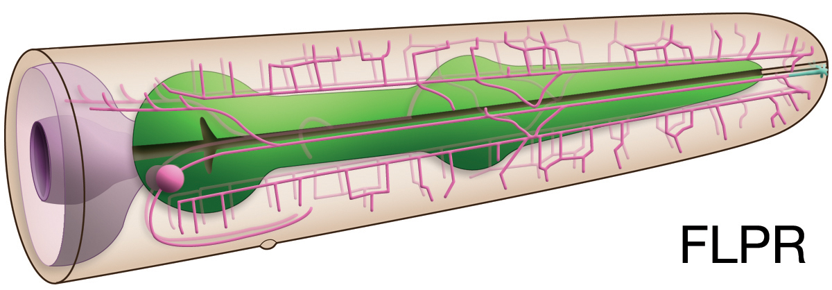
3D reconstruction of BAG and FLP cilia.
Both cilia are shown as associated with ILso process. Colors do not follow the WA color code (purple; BAG cilia, green; FLP cilia and dendritic endings, beige: ILso), ssTEM; serial section transmission electron micrography, TZ: transition zone. To expand, double click on the video, to return to original size, click "esc" (Doroquez et al., 2014)


 Click pictures for higher resolution images