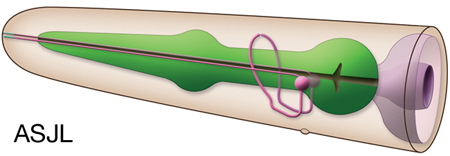
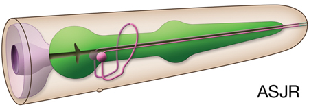
Type: Sensory neuron (dauer entry, electrosensory and photosensory) In MoW:ASJ Male Wiring Project:ASJLh,
ASJRh,
ASJLm,
ASJRm In Wormbase: ASJ, ASJL, ASJR Lineage:AB alpppppppa, AB praaappppa Location:Lateral ganglia of head Description:Amphid neurons, single (AsG) ciliated endings. Like all other amphid neurons, ASJ are born near the presumptive nose of the embryo during development. They then anchor a short projection there, after which the cell body migrates away, stretching the dendrite out behind it. This process is dependent on DEX-1 or DYF-7, secreted extracellular matrix proteins which act cooperatively for anchoring. In mutants lacking these proteins, the dendrite fails to anchor at the nose and is dragged along with the migrating cell body, giving rise to a short dendritic stub (Heiman and Shaham, 2010). Dendritic process takes up FITC. ASJ axon projects into the ventral cord by way of the same side amphid commissure and then grows into the nerve ring where it makes diverse synaptic connections in ring neuropil Neurotransmitter/ Neuropeptide:
- NLP-3; neuropeptide-like protein (Although earlier reports suggested ASJ might be glutamatergic, more recent studies showed it is not)
(Serrano-Saiz et al., 2013; Ohnishi et al., 2011; Nathoo et al., 2001) Innexin expression:
None yet reported, although described to have gap junctions in adult animals (MoW) Receptor expression:
- OSM-9; capsaicin receptor-like protein
- SRE-1; G protein-coupled, 7 TM protein
- DAF-11; transmembrane guanylyl cyclase (Birnby et al., 2000; Colbert et al., 1997; Troemel et al., 1995) Function:
- Control entry into dauer stage; ADF, ASI and ASG inhibit entry into dauer stage while ASJ and ASK promote dauer entry (Kim et al., 2009; Ouellett et al., 2008; Schackwitz et al., 1996). - Control exit from dauer stage; promotes dauer recovery (Bargmann and Horvitz, 1991).
- Lightsensation (350-470 nm range); when a flash of light is focused on the head of a worm moving forward, the animal halts and initiates reversals. Ablation of ASJ, AWB, ASK and ASH neurons together leads to a severe deficit in this head avoidance response while ablation of them individually or in different combinations does not yiled a significant defect suggesting functional redundancy
(Ward et al., 2008).
- Electrosensory navigation; C. elegans moves toward the negative pole of an electric field. Killing the ASJ or ASH neurons leads to significant disruption in electrotaxis while killing ASK, AWB or AWC has a weaker effect
(Gabel et al., 2007).
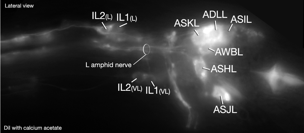
Click pictures for higher resolution images. You can also see DiI staining of ASJ in dorsoventral and lateral views.
Click here for larger version
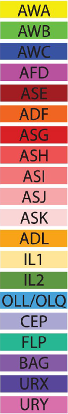
ASJL (AB alpppppppa) development in the embryo. Dorsal view. Bottom is left side of the embryo. Spheres indicate individual nuclei. Black sphere: ancestors of ASJL; dark grey spheres: apoptotic cells; other cells follow the WA color code (after they acquire specific cell or tissue identities). 0 min is fertilization. Click on the movie for higher resolution rendition (by A. Santella & Z. Bao).
Click here for larger version
ASJR (AB praaappppa) development in the embryo. Dorsal view. Bottom is left side of the embryo. Spheres indicate individual nuclei. Black sphere: ancestors of ASJR (since last ASJR ancestor has not yet gone through its final division, the black sphere seen at the end of this movie is still AB praaapppp); dark grey spheres: apoptotic cells; other cells follow the WA color code (after they acquire specific cell or tissue identities). 0 min is fertilization. Click on the movie for higher resolution rendition (by A. Santella & Z. Bao).
Click here for larger version
3D reconstruction of the anterior sensory endings (cilia and dendrites) from high resolution serial section transmission electron micrographs (ssTEMs). Bar 1 μm. Color code for the sensory endings is shown on the right-colors do not follow the WA color code. To expand, double click on the video, to return to original size, click "esc" (Doroquez et al., 2014)
3D
Click here for larger version
reconstruction of all amphid neuron cilia and associated socket and sheath cell processes. Modeled from serial section transmission electron micrographs (ssTEMs). Bar 1 μm. Color code for the sensory endings is shown on the left-colors do not follow the WA color code. To expand, double click on the video, to return to original size, click "esc" (Doroquez et al., 2014)


 Click pictures for higher resolution images. You can also see DiI staining of ASJ in