Neurotransmitter/ Neuropeptide:
- Glutamate
- FLP-6; FMRFamide-like neuropeptide
- NLP-7; neuropeptide-like peptide
- NLP-21; neuropeptide-like peptide
- Possibly PDF-1; pigment dispensing factor homolog
(Barrios et al, 2012; Ohnishi et al, 2011; Loer and Rand, 2022; Li and Kim, 2008; Rogers et al., 2003; Nathoo et al., 2001)
Innexin expression:
- CHE-7
- INX-1a
- INX-1b
- INX-2
- INX-7
- INX-10a
- INX-19
- UNC-7
(Altun et al., 2009; Bhattacharya et al., 2019; Chuang et al., 2007; Liu et al., 2006)
Receptor expression:
- GCY-8; transmembrane receptor guanylate cyclase (cGMP production in AFD is redundantly carried out by GCY-8, GCY-18 and GCY-23 and gcy-23, gcy-8, and gcy-18 triple mutants show a cryophilic or athermotactic phenotype(Inada et al., 2006)
- GCY-18; transmembrane receptor guanylate cyclase
- GCY-23; transmembrane receptor guanylate cyclase
- GCY-29; transmembrane receptor guanylate cyclase
- TMC-1; putative cation channel, salt-sensing receptor
(Wormbase; Chatzigeorgiou et al., 2013; Ortiz et al., 2006; Inada et al., 2006; Yu et al., 1997)
Function:
- Functions in thermotaxis: AFD neurons are the main thermosensors in C. elegans and laser ablation of the AFD pair makes most animals athermotactic (Ma and Shen, 2012; Beverly et al, 2011; Kuhara et al, 2008; Satterlee et al., 2004; Mori and Ohshima, 1995). After cultivation at a uniform temperature (Tc) with sufficient food, animals preferentially migrate to their cultivation temperature (Tc) when placed on a thermal gradient, and move isothermally at this temperature (Hedgecock and Russell, 1975). Animals sense and memorize their Tc by AFD (major thermosensory), AWC and ASI neurons (Beverly et al, 2011; Kuhara et al., 2008; Biron et al., 2008). This memory is plastic and can be reset upon cultivation at a different temperature (Hedgecock and Russell, 1975). To track isotherms, animals do not actively pursue isothermal alignment, but once serendipitously aligned along an isotherm (at T=Tc), they track by suppressing turns (Luo et al., 2006).The animal increases its reversal and turn frequency when it detects a rise in temperature and it moves back down the gradient toward Tc (negative thermotaxis). AFD neurons respond to thermal stimuli above Tc with continuous, graded calcium signals due to a Ca++ influx via cGMP-dependent TAX-2/TAX-4 cation channels. The AFD, AWC and ASI neurons may act in concert to increase turning rate when animals encounter higher temperatures (Biron et al., 2008). The thermal information is transmitted by AFD and AWC to AIY interneurons for information processing, and AIY neurons, in turn, transmit it to AIZ and RIA interneurons for further processing. It is suggested that activation of AFD neurons by warming above Tc induces the animal to reverse by inhibiting AIY since loss of AFD (by ablation) suppresses spontaneous reversals in an AIY-dependent manner, while loss of AIY (by ablation) increases spontaneous reversals (de Bono and Maricq 2005; Tsalik and Hobert, 2003). G-protein-coupled receptor (GPCR) SRTX-1 is required both in AFD and AWC for retaining normal isothermal tracking (Liu et al., 2012).
- In addition to sensing temperature within the viable range (~15-25oC), AFD neurons also take part in sensing noxious temperatures (thermonociception) (~35-38oC). Noxious temperatures induce a temperature avoidance response with reflex-like escape reaction. FLP neurons in the head and PHC neurons in the tail also act as thermonociceptive neurons. This avoidance response requires the cell autonomous function of TAX-2, TAX-4 in AFD, and the heat- and capsaicin-sensitive transient receptor potential vanilloid (TRPV) channels, OCR-2/OSM-9 in the FLP and PHC neurons. In sensing ambient temperature, cGMP production in AFD is through GCY-23, GCY-8 and GCY-18, while for noxious heat sensation cGMP production in AFD is mainly through GCY-12. Noxious temperature avoidance via AFD involves AIB interneuron (Liu et al., 2012).
- Functions in locomotion: Laser ablations of AFD cause hyporeversal phenotype (Tsalik and Hobert, 2003).
- AFD, BAG and ASE are primary CO2 sensors, while oxygen-sensing neurons AQR, PQR and URX are also weakly CO2 responsive. AFD and BAG neurons together stimulate turning when CO2 rises and inhibit turning when CO2 falls (Bretscher et al., 2011).
- May be involved in social feeding since disruption of tax-2 expression in AFD, AQR, ASE and BAG neurons disrupts social feeding (Coates and de Bono, 2002).
Reporters:
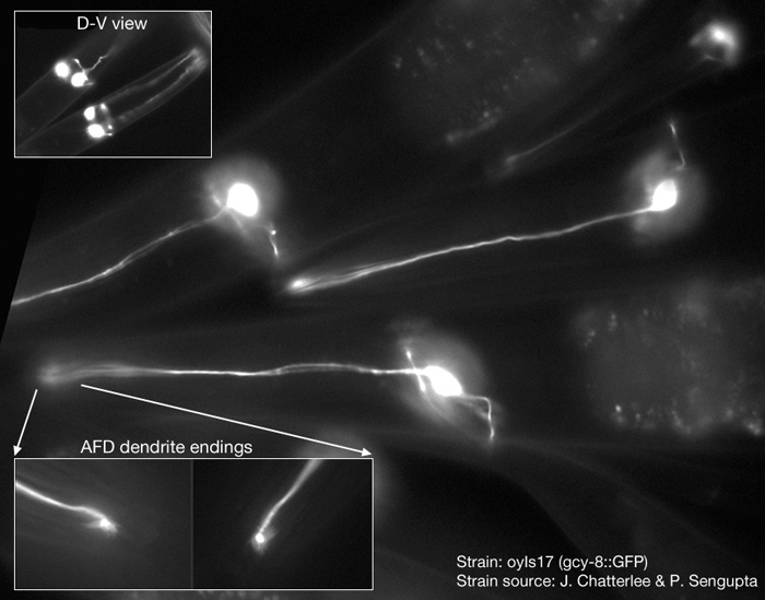
- At CGC: PY1157 oyls17 [gcy-8p::GFP + lin-15(+)].
- Other:
|


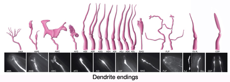
 Click pictures for higher resolution images