|
Usage Guide:
General Guides
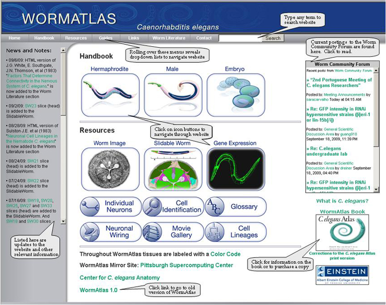
Front Page:
The front page of WormAtlas contains lots of information and links to help navigate through the website. The top menu bar is found on every page of the website. See picture below for detailed information.
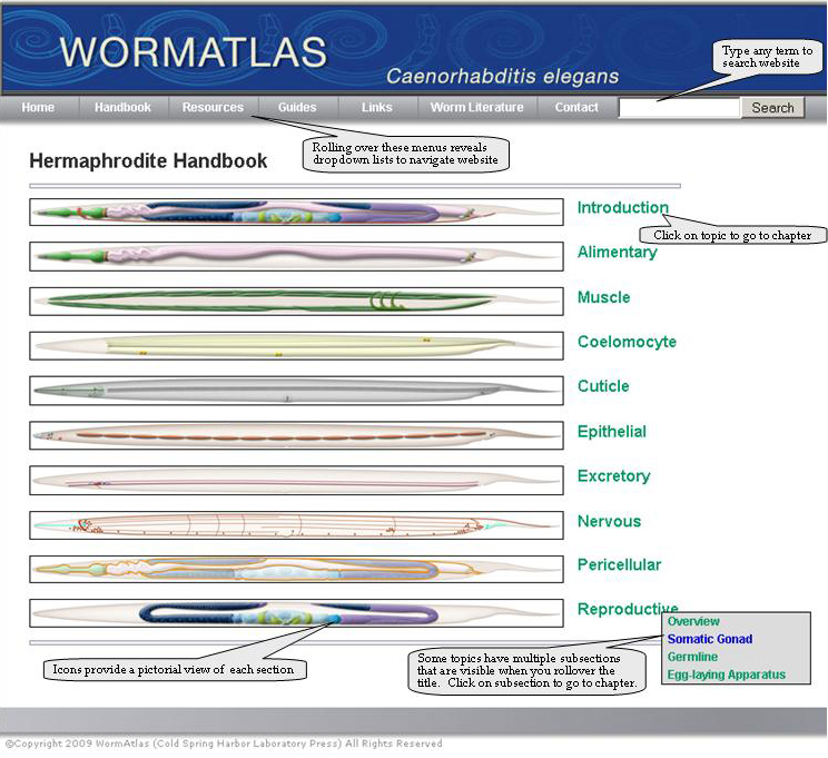
Handbook Contents Page:
Clicking on the hermaphrodite or male handbook icons or using the drop down menus takes you to a table of contents page. Each tissue type is featured and illustrated with a picture. On the right side of each picture are the names of each section. Some sections only have one chapter and for those you can click on the name and you will go directly to the chapter text. Other sections have subsections and those can be found by rolling over the name and a new list of clickable subsections will appear. Clicking on these titles will take you to the appropriate chapter.
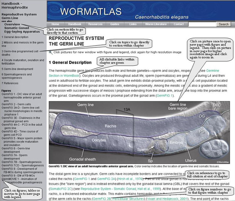
Chapter Pages:
Each chapter is organized with a table of contents on the left-hand side. You can click on the headings within the table of contents to go directly to those sections within the chapter. Clicking on the one of the names of the figures or tables within the list will take you to a new page with the figure and legend. (Note that in the male handbook, clicking on the figure number will take you to the figure within the text of the chapter and you cannot click on the pictures to go to higher resolution images.)
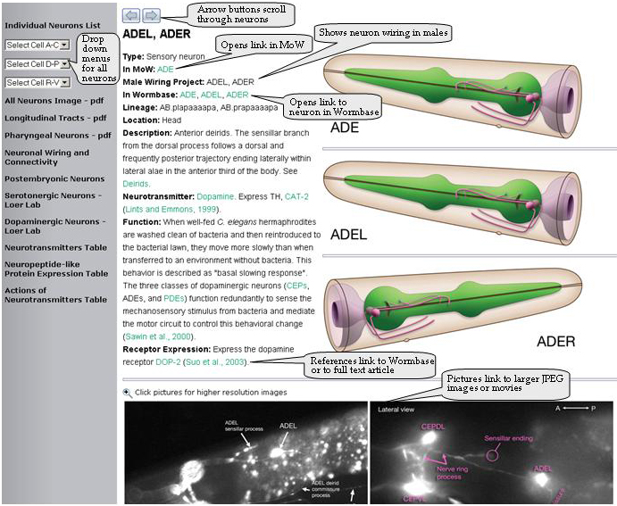
Individual Neuron Pages:
On each individual neuron page you can open several links to view all the information regarding that specific neuron in Wormbase, Mind of the Worm or at the Male Wiring Project. Information about each neuron can be found in the description, neurotransmitter, function and receptor expression categories. Within the text, there are also links that take you to more relevant information in WormBook or in other sections of WormAtlas. Clicking on references will open citations in WormBase or to the full text of the article if it is available. Clicking on the expression pattern pictures opens a higher resolution image file. Additionally, each neuron page has a left hand sidebar that allows you to select other neurons by alphabetical drop down menu. Other images and related neuron links are also found in this side bar. Use the arrows at the top of the page to quickly scroll through the neuron pages in alphabetical order.
Neuron Guides
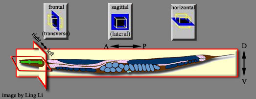
Image Planes:
The planes of image sections used in WormAtlas are as shown below:
Neuron images:
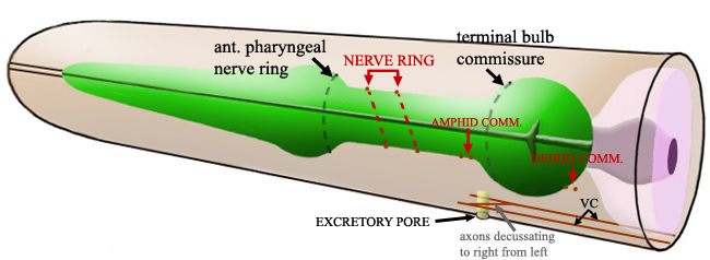
In images of head neurons, burgundy dots refer to the locations of nerve ring, amphid commissure and deirid commissure respectively from anterior to posterior. Straight burgundy lines below pharynx and intestine refer to the right and left sides of the ventral cord as shown below:
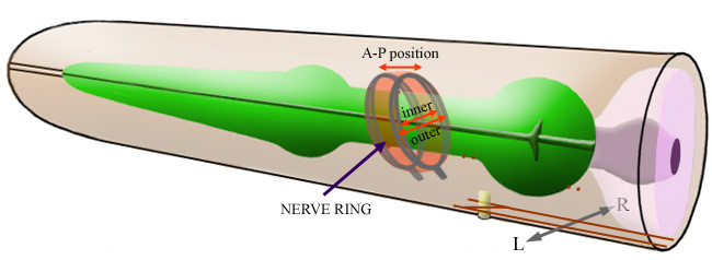
The radius of the circle an axon makes in the nerve ring defines the location of this axon in ring neuropile. The smallest radius belongs to those axons traveling in the inner sections of the ring neuropile, while the largest radius belongs to those traveling in the outer section. Middle range radii belong to those traveling in the middle of the ring neuropile.
The placement of an axon in the antero-posterior alignment reflects the position of that axon in A-P axis in the ring neuropile.
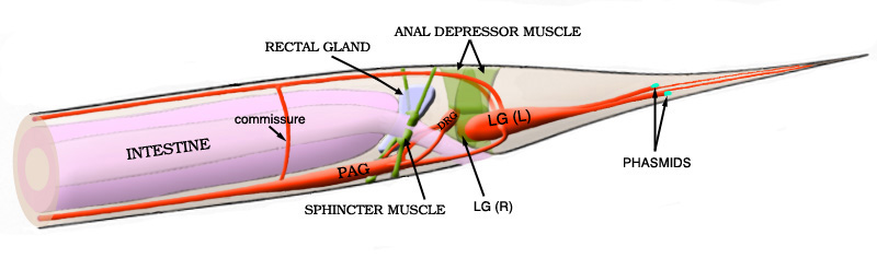
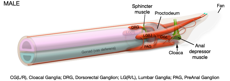
The tail neurons are placed in the tail ganglia shown below:
|
