|
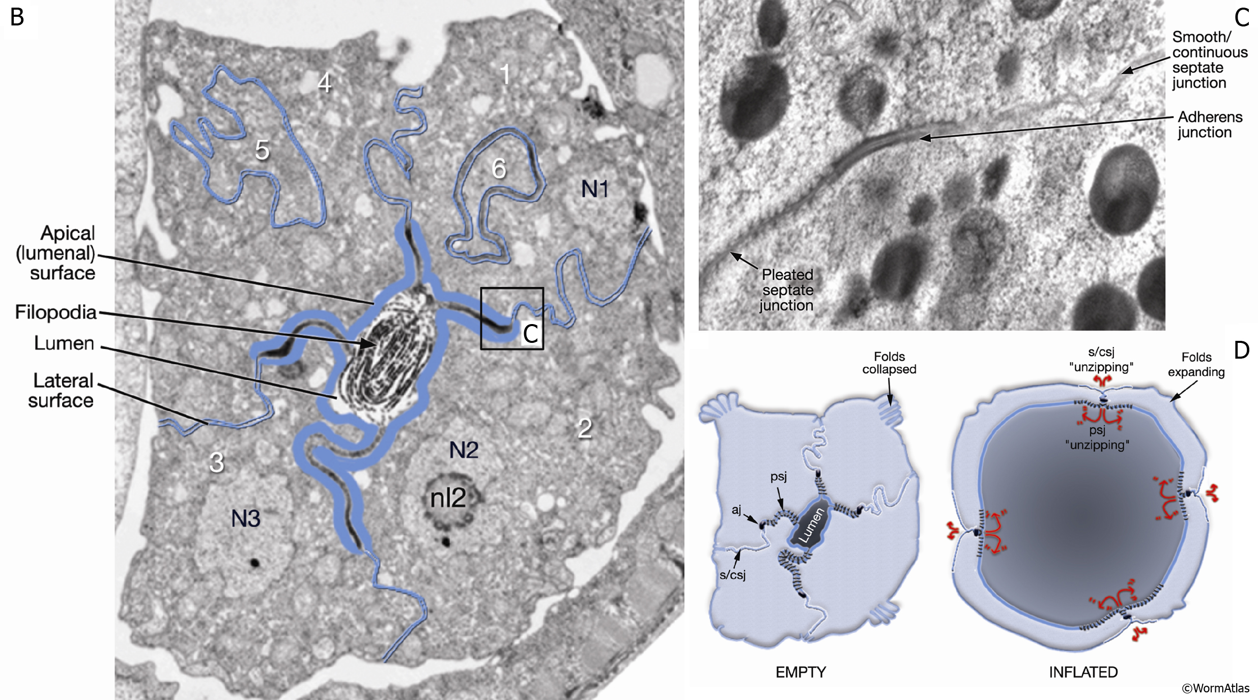
SomaticFIG 9B-D: Cell junctions of the spermatheca.
B. TEM, transverse section of a late L4 hermaphrodite spermatheca (before the first ovulation). The number of cells that make up the spermathecal wall is indicated. The lumenal (apical) surface (heavy blue lines) bears filopodia, pleated septate junctions, and adherens junctions. The lateral cell borders (thin blue lines) contain smooth/continuous septate junctions and gap junctions (see C). (N) Nucleus; (nl) nucleolus. (Image source: VS8/1 [MRC] 4935-3.)
C. TEM, transverse section showing a high-magnification view of junction types from the areas indicated by the box in B. (Image source: Hall archive.)
D. Schematic of the spermatheca (transverse view) showing the possible roles for junction types in spermatheca function (see text). (aj) Adherens junction; (psj) pleated septate junction; (s/csj) smooth/continuous septate junction.
Click on picture for full resolution image.
|