|
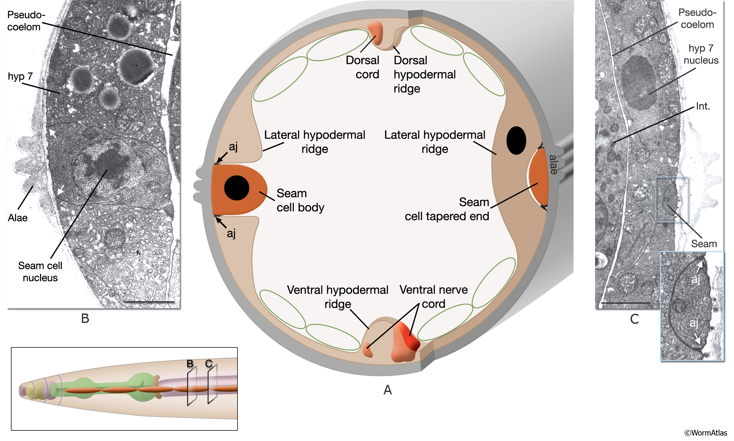
SeamFIG 2: Seam cells are embedded in hypodermis.
A. The hypodermis is composed of four major longitudinal ridges (ventral, dorsal, and L/R lateral) that are joined circumferentially by thin sheets of cytoplasm. At each original cell body of the seam syncytium, where the nuclei are positioned, the seam completely interrupts hyp 7 (section through B in inset), whereas at the ends of the cells, the seam is so thin that hyp 7 runs behind it, enfolding it completely (section through C in inset). At its apical surface, seam makes adherens junctions (aj) to the hypodermis throughout its length. (Green circles) Muscle quadrants.
B. TEM, transverse section representing an area through the seam-cell body in an adult animal. The body of the seam is lodged between the ventral and dorsal parts of the lateral hypodermal ridge. Its basal surface is surrounded by the pseudocoelom, although it is separated from the pseudocoelom by a basal lamina. Its apical surface is in complete register with cuticular alae. (White arrows) Adherens junctions. (Image source: B140 [Hall] A578.) Bar, 1 μm.
C. TEM, transverse section representing area through tip of seam cell in an adult animal. At this level, hyp 7 completely wraps around the animal, running behind the seam. The lateral nuclei of the hyp7 syncytium are located on the ventral or dorsal side of the seam at this level. (Int.) Intestine. (Image source: [Hall] 8396-1.) Bar, 1 μm.
Click on picture for full resolution image.
|