|
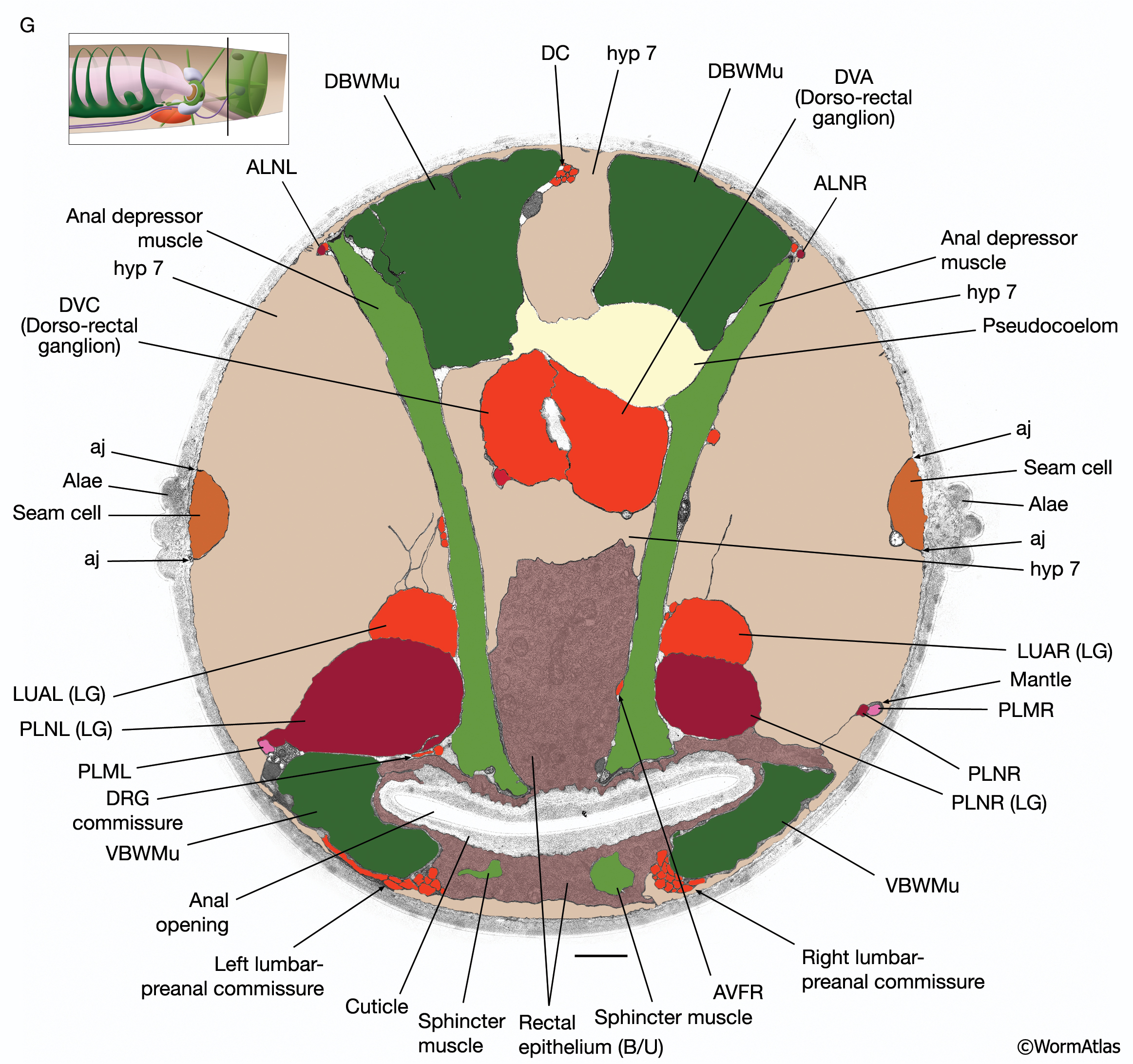
RectFIG 3G: The rectum.
Transverse pseudocolored TEM images of the adult rectum. The K cell has divided and the Y cell has become PDA (not shown). Hyp 12 becomes part of hyp7. (DC) Dorsal cord; (LG) lumbar ganglia; (aj) adherens junction; (DBWMu and VBWMu) dorsal and ventral body wall muscles, respectively. FIG 3F shows the middle of the rectum, whereas G is closer to the anal opening. (Image source: [Hall] B140B [dark gray areas are gaps in the original TEM montage]. Bar, 1 μm.
See also RectFIG 3A-E and 3F.
Click on picture for full resolution image.
|